 Movie
Movie Controller

[English] 日本語
 Yorodumi
- PDB-1h5z: CYTOCHROME P450 14 ALPHA-STEROL DEMETHYLASE (CYP51) FROM MYCOBACT... -
+ Open data

- Basic information
| Entry | Database: PDB / ID: 1h5z | ||||||
|---|---|---|---|---|---|---|---|
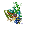
| Title | CYTOCHROME P450 14 ALPHA-STEROL DEMETHYLASE (CYP51) FROM MYCOBACTERIUM TUBERCULOSIS IN FERRIC LOW-SPIN STATE | ||||||
 Components Components | CYTOCHROME P450 51 | ||||||
 Keywords Keywords | OXIDOREDUCTASE / CYTOCHROME P450 / 14 ALPHA-STEROL DEMETHYLASE / FERRIC LOW-SPIN / STEROL BIOSYNTHESIS / MONOOXYGENASE / ELECTRON TRANSPORT | ||||||
| Function / homology |  Function and homology information Function and homology informationsterol 14alpha-demethylase (ferredoxin) / steroid 7-alpha-hydroxylase activity / oxysterol 7-alpha-hydroxylase activity / sterol 14alpha-demethylase / sterol 14-demethylase activity / sterol biosynthetic process / bile acid biosynthetic process / sterol metabolic process / oxidoreductase activity, acting on paired donors, with incorporation or reduction of molecular oxygen / cholesterol homeostasis ...sterol 14alpha-demethylase (ferredoxin) / steroid 7-alpha-hydroxylase activity / oxysterol 7-alpha-hydroxylase activity / sterol 14alpha-demethylase / sterol 14-demethylase activity / sterol biosynthetic process / bile acid biosynthetic process / sterol metabolic process / oxidoreductase activity, acting on paired donors, with incorporation or reduction of molecular oxygen / cholesterol homeostasis / monooxygenase activity / oxidoreductase activity / iron ion binding / heme binding / cytosol / cytoplasmSimilarity search - Function | ||||||
| Biological species |   MYCOBACTERIUM TUBERCULOSIS (bacteria) MYCOBACTERIUM TUBERCULOSIS (bacteria) | ||||||
| Method | X-RAY DIFFRACTION / MOLECULAR REPLACEMENT / Resolution: 2.05 Å | ||||||
 Authors Authors | Podust, L.M. / Arase, M. / Waterman, M.R. | ||||||
 Citation Citation | Journal: Structure / Year: 2004 Title: Estriol Bound and Ligand-Free Structures of Sterol 14Alpha-Demethylase. Authors: Podust, L.M. / Yermalitskaya, L.V. / Lepesheva, G.I. / Podust, V.N. / Dalmasso, E.A. / Waterman, M.R. #1: Journal: J.Inorg.Biochem. / Year: 2001 Title: Substrate Recognition Sites in 14Alpha-Sterol Demethylase from Comparative Analysis of Amino Acid Sequences and X-Ray Structure of Mycobacterium Tuberculosis Cyp51. Authors: Podust, L.M. / Stojan, J. / Poulos, T.L. / Waterman, M.R. #2: Journal: Proc.Natl.Acad.Sci.USA / Year: 2001Title: Crystal Structure of Cytochrome P450 14Alpha -Sterol Demethylase (Cyp51) from Mycobacterium Tuberculosis in Complex with Azole Inhibitors. Authors: Podust, L.M. / Poulos, T.L. / Waterman, M.R. | ||||||
| History |
|
- Structure visualization
| Structure viewer | Molecule: MolmilJmol/JSmol |
|---|
- Downloads & links
-Download
| PDBx/mmCIF format | 1h5z.cif.gz | 108.5 KB | Display | PDBx/mmCIF format |
|---|---|---|---|---|
| PDB format | pdb1h5z.ent.gz | 81.1 KB | Display | PDB format |
| PDBx/mmJSON format | 1h5z.json.gz | Tree view | PDBx/mmJSON format | |
| Others |  Other downloads Other downloads |
-Validation report
| Arichive directory | https://data.pdbj.org/pub/pdb/validation_reports/h5/1h5zftp://data.pdbj.org/pub/pdb/validation_reports/h5/1h5z | HTTPS FTP |
|---|
-Related structure data
| Related structure data |  1x8vC  1e9xS S: Starting model for refinement C: citing same article ( |
|---|---|
| Similar structure data |


-Links
 PDBj
PDBj


- Assembly
| Deposited unit | 
| ||||||||
|---|---|---|---|---|---|---|---|---|---|
| 1 |
| ||||||||
| Unit cell |
|
-Components
| #1: Protein | / CYP51 / STEROL 14-ALPHA DEMETHYLASE / LANOSTEROL 14-ALPHA DEMETHYLASE / P450-14DM Mass: 51499.332 Da / Num. of mol.: 1 Source method: isolated from a genetically manipulated source Details: CYS 394 BINDS HEME IRON / Source: (gene. exp.) MYCOBACTERIUM TUBERCULOSIS (bacteria) / Cellular location: CYTOPLASM / Gene: CYP51, RV0764C, MTCY369.09C / Plasmid: PET17B / Production host: ESCHERICHIA COLI (E. coli) / Strain (production host): HMS174References: UniProt: P77901, UniProt: P9WPP9*PLUS, sterol 14alpha-demethylase |
|---|---|
| #2: Chemical | ChemComp-HEM / Heme B  Mass: 616.487 Da / Num. of mol.: 1 / Source method: obtained synthetically / Formula: C34H32FeN4O4 Mass: 616.487 Da / Num. of mol.: 1 / Source method: obtained synthetically / Formula: C34H32FeN4O4 |
| #3: Chemical | ChemComp-FE2 /   Mass: 55.845 Da / Num. of mol.: 1 / Source method: obtained synthetically / Formula: Fe Mass: 55.845 Da / Num. of mol.: 1 / Source method: obtained synthetically / Formula: Fe |
| #4: Water | ChemComp-HOH / Water Mass: 18.015 Da / Num. of mol.: 280 / Source method: isolated from a natural source / Formula: H2O Mass: 18.015 Da / Num. of mol.: 280 / Source method: isolated from a natural source / Formula: H2O |
| Sequence details | FOUR HIS RESIDUES WERE ENGINEERED |
-Experimental details
-Experiment
| Experiment | Method: X-RAY DIFFRACTION / Number of used crystals: 1 |
|---|
- Sample preparation
| Crystal | Density Matthews: 2.132 Å3/Da / Density % sol: 41 % | |||||||||||||||||||||||||||||||||||||||||||||||||
|---|---|---|---|---|---|---|---|---|---|---|---|---|---|---|---|---|---|---|---|---|---|---|---|---|---|---|---|---|---|---|---|---|---|---|---|---|---|---|---|---|---|---|---|---|---|---|---|---|---|---|
| Crystal grow | Temperature: 295 K / pH: 6.5 / Details: 20% PEG 4000, 0.1 M NA CACODYLATE, PH 6.5, 22 C | |||||||||||||||||||||||||||||||||||||||||||||||||
| Crystal grow | *PLUS pH: 7.5 / Method: vapor diffusion, hanging drop | |||||||||||||||||||||||||||||||||||||||||||||||||
| Components of the solutions | *PLUS
|
-Data collection
| Diffraction | Mean temperature: 100 K |
|---|---|
| Diffraction source | Source: ROTATING ANODE / Type: RIGAKU RU200 / Wavelength: 1.5418 |
| Detector | Type: RIGAKU-MSC / Detector: IMAGE PLATE / Date: Mar 22, 2001 / Details: MIRROR |
| Radiation | Monochromator: NI / Protocol: SINGLE WAVELENGTH / Monochromatic (M) / Laue (L): M / Scattering type: x-ray |
| Radiation wavelength | Wavelength: 1.5418 Å / Relative weight: 1 |
| Reflection | Resolution: 2.05→40 Å / Num. obs: 25277 / % possible obs: 99.6 % / Observed criterion σ(I): 0 / Redundancy: 4.1 % / Biso Wilson estimate: 19.1 Å2 / Rsym value: 0.105 / Net I/σ(I): 10.6 |
| Reflection shell | Resolution: 2.05→2.12 Å / Redundancy: 3.1 % / Mean I/σ(I) obs: 3.1 / Rsym value: 0.457 / % possible all: 90.9 |
| Reflection | *PLUS Highest resolution: 2.05 Å / Lowest resolution: 40 Å / % possible obs: 90.6 % / Redundancy: 4.1 % / Num. measured all: 103288 / Rmerge(I) obs: 0.105 |
| Reflection shell | *PLUS % possible obs: 90.9 % / Redundancy: 3.1 % / Rmerge(I) obs: 0.457 / Mean I/σ(I) obs: 3.1 |
- Processing
| Software |
| ||||||||||||||||||||||||||||||||||||||||||||||||||||||||||||||||||||||||||||||||
|---|---|---|---|---|---|---|---|---|---|---|---|---|---|---|---|---|---|---|---|---|---|---|---|---|---|---|---|---|---|---|---|---|---|---|---|---|---|---|---|---|---|---|---|---|---|---|---|---|---|---|---|---|---|---|---|---|---|---|---|---|---|---|---|---|---|---|---|---|---|---|---|---|---|---|---|---|---|---|---|---|---|
| Refinement | Method to determine structure: MOLECULAR REPLACEMENT Starting model: PDB ENTRY 1E9X Resolution: 2.05→35.51 Å / Rfactor Rfree error: 0.005 / Data cutoff high absF: 203124.28 / Isotropic thermal model: RESTRAINED / Cross valid method: THROUGHOUT / σ(F): 0 / Details: HIS TAG ON THE C-TERMINAL
| ||||||||||||||||||||||||||||||||||||||||||||||||||||||||||||||||||||||||||||||||
| Solvent computation | Solvent model: FLAT MODEL / Bsol: 52.054 Å2 / ksol: 0.375135 e/Å3 | ||||||||||||||||||||||||||||||||||||||||||||||||||||||||||||||||||||||||||||||||
| Displacement parameters | Biso mean: 27 Å2
| ||||||||||||||||||||||||||||||||||||||||||||||||||||||||||||||||||||||||||||||||
| Refine analyze |
| ||||||||||||||||||||||||||||||||||||||||||||||||||||||||||||||||||||||||||||||||
| Refinement step | Cycle: LAST / Resolution: 2.05→35.51 Å
| ||||||||||||||||||||||||||||||||||||||||||||||||||||||||||||||||||||||||||||||||
| Refine LS restraints |
| ||||||||||||||||||||||||||||||||||||||||||||||||||||||||||||||||||||||||||||||||
| LS refinement shell | Resolution: 2.05→2.18 Å / Rfactor Rfree error: 0.016 / Total num. of bins used: 6
| ||||||||||||||||||||||||||||||||||||||||||||||||||||||||||||||||||||||||||||||||
| Xplor file |
| ||||||||||||||||||||||||||||||||||||||||||||||||||||||||||||||||||||||||||||||||
| Refinement | *PLUS Lowest resolution: 40 Å | ||||||||||||||||||||||||||||||||||||||||||||||||||||||||||||||||||||||||||||||||
| Solvent computation | *PLUS | ||||||||||||||||||||||||||||||||||||||||||||||||||||||||||||||||||||||||||||||||
| Displacement parameters | *PLUS | ||||||||||||||||||||||||||||||||||||||||||||||||||||||||||||||||||||||||||||||||
| Refine LS restraints | *PLUS
|
