 Movie
Movie Controller

[English] 日本語
 Yorodumi
- PDB-1gpm: ESCHERICHIA COLI GMP SYNTHETASE COMPLEXED WITH AMP AND PYROPHOSPHATE -
+ Open data

- Basic information
| Entry | Database: PDB / ID: 1gpm | ||||||
|---|---|---|---|---|---|---|---|
| Title | ESCHERICHIA COLI GMP SYNTHETASE COMPLEXED WITH AMP AND PYROPHOSPHATE | ||||||
 Components Components | GMP SYNTHETASE GMP synthase GMP synthase | ||||||
 Keywords Keywords | TRANSFERASE (GLUTAMINE AMIDOTRANSFERASE) / CLASS I GLUTAMINE AMIDOTRANSFERASE / N-TYPE ATP PYROPHOSPHATASE | ||||||
| Function / homology |  Function and homology informationGMP synthase activity / GMP synthase (glutamine-hydrolyzing) activity / GMP synthase (glutamine-hydrolysing) / pyrophosphatase activity / GMP biosynthetic process / glutamine metabolic process / ATP binding / identical protein binding / cytosol Function and homology informationGMP synthase activity / GMP synthase (glutamine-hydrolyzing) activity / GMP synthase (glutamine-hydrolysing) / pyrophosphatase activity / GMP biosynthetic process / glutamine metabolic process / ATP binding / identical protein binding / cytosolSimilarity search - Function | ||||||
| Biological species |  Escherichia coli (E. coli) Escherichia coli (E. coli) | ||||||
| Method | X-RAY DIFFRACTION / SYNCHROTRON / Resolution: 2.2 Å | ||||||
 Authors Authors | Tesmer, J.J.G. | ||||||
 Citation Citation | Journal: Nat.Struct.Biol. / Year: 1996 Title: The crystal structure of GMP synthetase reveals a novel catalytic triad and is a structural paradigm for two enzyme families. Authors: Tesmer, J.J. / Klem, T.J. / Deras, M.L. / Davisson, V.J. / Smith, J.L. #1: Journal: Proteins / Year: 1994Title: Preliminary X-Ray Analysis of Escherichia Coli Gmp Synthetase: Determination of Anomalous Scattering Factors for a Cysteinyl Mercury Derivative Authors: Tesmer, J.J.G. / Stemmler, T.L. / Penner-Hahn, J.E. / Davisson, V.J. / Smith, J.L. | ||||||
| History |
| ||||||
| Remark 650 | HELIX HELIX DETERMINATION METHOD: RESIDUES FORMING SECONDARY STRUCTURE HAVE BACKBONE H-BONDS < 3.4 ...HELIX HELIX DETERMINATION METHOD: RESIDUES FORMING SECONDARY STRUCTURE HAVE BACKBONE H-BONDS < 3.4 ANGSTROMS HELIX_ID: 5A,LAST 2 RESIDUES FORM PI H-BONDS. HELIX_ID: 8A,LAST 2 RESIDUES FORM PI H-BONDS. HELIX_ID: 5B,LAST 2 RESIDUES FORM PI H-BONDS. HELIX_ID: 8B,LAST 2 RESIDUES FORM PI H-BONDS. HELIX_ID: 5C,LAST 2 RESIDUES FORM PI H-BONDS. HELIX_ID: 8C,LAST 2 RESIDUES FORM PI H-BONDS. HELIX_ID: 5D,LAST 2 RESIDUES FORM PI H-BONDS. HELIX_ID: 8D,LAST 2 RESIDUES FORM PI H-BONDS. | ||||||
| Remark 700 | SHEET S1A, S1B, S1C, S1D: DETERMINATION METHOD: RESIDUES FORMING SECONDARY STRUCTURE HAVE BACKBONE ...SHEET S1A, S1B, S1C, S1D: DETERMINATION METHOD: RESIDUES FORMING SECONDARY STRUCTURE HAVE BACKBONE H-BONDS < 3.4 ANGSTROMS; S1A AND S2A, S1B AND S2B, S1C AND S2C, S1D AND S2D ARE CONTINUOUS, BUT THE OVERALL FOLD OF THE DOMAIN SUGGESTS THAT THEY ARE BETTER DESCRIBED AS DISTINCT SHEETS. THE FIRST STRAND IN S2A, S2B, S2C, S2D WOULD BE THE NEXT STRAND IN S1A S1B, S1C, S1D, RESPECTIVELY. S3A, S3B, S3C, S3D: BETA RIBBON. THE DIMERIZATION DOMAIN FOR CHAINS A AND C CONTAINS A SHEET WITH TWO BIFURCATED STRANDS, AS DOES THE DIMERIZATION DOMAIN FOR CHAINS B AND D. THESE SHEETS ARE PRESENTED AS SHEETS SAC AND TAC FOR CHAINS A AND C AND SHEETS SBD AND TBD FOR CHAINS B AND D. SHEETS SAC AND TAC DIFFER IN CHAINS 3 AND 4 AS DO SHEETS SBD AND TBD. SBD: DIMERIZATION DOMAIN. ATOM_NAME: () () CYS A 86 () CYS 86 HAS A STRAINED BACKBONE CONFORMATION (PHI=55, PSI= -110) TYPICAL OF "NUCLEOPHILE ELBOWS" FOUND IN THE A/B HYDROLASES. THE RESIDUE IS BOTH A MEMBER OF SHEET S1A AND HELIX 4A. ATOM_NAME: () () CYS B 86 () CYS 86 HAS A STRAINED BACKBONE CONFORMATION (PHI=55, PSI= -110) TYPICAL OF "NUCLEOPHILE ELBOWS" FOUND IN THE A/B HYDROLASES. THE RESIDUE IS BOTH A MEMBER OF SHEET S1B AND HELIX 4B. ATOM_NAME: () () CYS C 86 () CYS 86 HAS A STRAINED BACKBONE CONFORMATION (PHI=55, PSI= -110) TYPICAL OF "NUCLEOPHILE ELBOWS" FOUND IN THE A/B HYDROLASES. THE RESIDUE IS BOTH A MEMBER OF SHEET S1C AND HELIX 4C. ATOM_NAME: () () CYS D 86 () CYS 86 HAS A STRAINED BACKBONE CONFORMATION (PHI=55, PSI= -110) TYPICAL OF "NUCLEOPHILE ELBOWS" FOUND IN THE A/B HYDROLASES. THE RESIDUE IS BOTH A MEMBER OF SHEET S1D AND HELIX 4D. |
- Structure visualization
| Structure viewer | Molecule: MolmilJmol/JSmol |
|---|
- Downloads & links
-Download
| PDBx/mmCIF format | 1gpm.cif.gz | 418 KB | Display | PDBx/mmCIF format |
|---|---|---|---|---|
| PDB format | pdb1gpm.ent.gz | 340.8 KB | Display | PDB format |
| PDBx/mmJSON format | 1gpm.json.gz | Tree view | PDBx/mmJSON format | |
| Others |  Other downloads Other downloads |
-Validation report
| Arichive directory | https://data.pdbj.org/pub/pdb/validation_reports/gp/1gpmftp://data.pdbj.org/pub/pdb/validation_reports/gp/1gpm | HTTPS FTP |
|---|
-Related structure data
| Similar structure data |
|---|


-Links
 PDBj
PDBj


- Assembly
| Deposited unit | 
| ||||||||
|---|---|---|---|---|---|---|---|---|---|
| 1 |
| ||||||||
| Unit cell |
| ||||||||
| Atom site foot note | 1: CIS PROLINE - PRO A 518 / 2: CIS PROLINE - PRO A 519 / 3: CIS PROLINE - PRO B 518 / 4: CIS PROLINE - PRO B 519 / 5: CIS PROLINE - PRO C 518 / 6: CIS PROLINE - PRO C 519 / 7: CIS PROLINE - PRO D 518 / 8: CIS PROLINE - PRO D 519 |
-Components
-Protein , 1 types, 4 molecules ABCD
| #1: Protein | GMP synthase / XMP AMINASE Mass: 58750.043 Da / Num. of mol.: 4 Source method: isolated from a genetically manipulated source Source: (gene. exp.) Escherichia coli (E. coli) / Strain: K12 / Description: TAC PROMOTER / Gene: GUAA / Plasmid: PGUAA-TAC / Gene (production host): GUAA / Production host: Escherichia coli (E. coli)References: UniProt: P04079, GMP synthase (glutamine-hydrolysing) |
|---|
-Non-polymers , 6 types, 809 molecules 


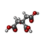


| #2: Chemical | ChemComp-PO4 / Phosphate Mass: 94.971 Da / Num. of mol.: 4 / Source method: obtained synthetically / Formula: PO4 Mass: 94.971 Da / Num. of mol.: 4 / Source method: obtained synthetically / Formula: PO4#3: Chemical | ChemComp-POP / Pyrophosphate Mass: 175.959 Da / Num. of mol.: 4 / Source method: obtained synthetically / Formula: H2O7P2 Mass: 175.959 Da / Num. of mol.: 4 / Source method: obtained synthetically / Formula: H2O7P2#4: Chemical | ChemComp-AMP / Adenosine monophosphate Mass: 347.221 Da / Num. of mol.: 4 / Source method: obtained synthetically / Formula: C10H14N5O7P / Comment: AMP*YM Mass: 347.221 Da / Num. of mol.: 4 / Source method: obtained synthetically / Formula: C10H14N5O7P / Comment: AMP*YM#5: Chemical | ChemComp-CIT / Citric acid Mass: 192.124 Da / Num. of mol.: 4 / Source method: obtained synthetically / Formula: C6H8O7 Mass: 192.124 Da / Num. of mol.: 4 / Source method: obtained synthetically / Formula: C6H8O7#6: Chemical |  Mass: 24.305 Da / Num. of mol.: 3 / Source method: obtained synthetically / Formula: Mg Mass: 24.305 Da / Num. of mol.: 3 / Source method: obtained synthetically / Formula: Mg#7: Water | ChemComp-HOH / | WaterMass: 18.015 Da / Num. of mol.: 790 / Source method: isolated from a natural source / Formula: H2O |
|---|
-Details
| Sequence details | N-TERMINAL SEQUENCING |
|---|
-Experimental details
-Experiment
| Experiment | Method: X-RAY DIFFRACTION |
|---|
- Sample preparation
| Crystal |
| ||||||||||||||||||||||||||||||||||||||||||||||||||||||||||||||||||
|---|---|---|---|---|---|---|---|---|---|---|---|---|---|---|---|---|---|---|---|---|---|---|---|---|---|---|---|---|---|---|---|---|---|---|---|---|---|---|---|---|---|---|---|---|---|---|---|---|---|---|---|---|---|---|---|---|---|---|---|---|---|---|---|---|---|---|---|
| Crystal grow | pH: 5 / Details: pH 5.0 | ||||||||||||||||||||||||||||||||||||||||||||||||||||||||||||||||||
| Crystal grow | *PLUS Temperature: 20 ℃ / Method: vapor diffusion, sitting drop | ||||||||||||||||||||||||||||||||||||||||||||||||||||||||||||||||||
| Components of the solutions | *PLUS
|
-Data collection
| Diffraction |
| |||||||||||||||||||||
|---|---|---|---|---|---|---|---|---|---|---|---|---|---|---|---|---|---|---|---|---|---|---|
| Diffraction source |
| |||||||||||||||||||||
| Detector |
| |||||||||||||||||||||
| Radiation | Monochromatic (M) / Laue (L): M / Scattering type: x-ray | |||||||||||||||||||||
| Radiation wavelength |
| |||||||||||||||||||||
| Reflection | Resolution: 2.2→50 Å / Num. obs: 105224 / % possible obs: 84.5 % / Observed criterion σ(I): 0 / Redundancy: 4.2 % / Rmerge(I) obs: 0.053 | |||||||||||||||||||||
| Reflection | *PLUS % possible obs: 841.4 % / Rmerge(I) obs: 0.059 | |||||||||||||||||||||
| Reflection shell | *PLUS % possible obs: 48.4 % / Rmerge(I) obs: 0.149 / Mean I/σ(I) obs: 8.7 |

- Processing
| Software |
| ||||||||||||||||||||||||||||||||||||||||||||||||||||||||||||
|---|---|---|---|---|---|---|---|---|---|---|---|---|---|---|---|---|---|---|---|---|---|---|---|---|---|---|---|---|---|---|---|---|---|---|---|---|---|---|---|---|---|---|---|---|---|---|---|---|---|---|---|---|---|---|---|---|---|---|---|---|---|
| Refinement | Resolution: 2.2→6 Å /
| ||||||||||||||||||||||||||||||||||||||||||||||||||||||||||||
| Displacement parameters | Biso mean: 35.3 Å2 | ||||||||||||||||||||||||||||||||||||||||||||||||||||||||||||
| Refinement step | Cycle: LAST / Resolution: 2.2→6 Å
| ||||||||||||||||||||||||||||||||||||||||||||||||||||||||||||
| Refine LS restraints |
| ||||||||||||||||||||||||||||||||||||||||||||||||||||||||||||
| Software | *PLUS Name: X-PLOR / Classification: refinement | ||||||||||||||||||||||||||||||||||||||||||||||||||||||||||||
| Refinement | *PLUS | ||||||||||||||||||||||||||||||||||||||||||||||||||||||||||||
| Solvent computation | *PLUS | ||||||||||||||||||||||||||||||||||||||||||||||||||||||||||||
| Displacement parameters | *PLUS | ||||||||||||||||||||||||||||||||||||||||||||||||||||||||||||
| Refine LS restraints | *PLUS
|
