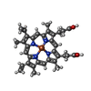
cytochrome o ubiquinol oxidase complex / oxidoreduction-driven active transmembrane transporter activity / ubiquinol oxidase (H+-transporting) / cytochrome bo3 ubiquinol oxidase activity / aerobic electron transport chain / oxidoreductase activity, acting on diphenols and related substances as donors, oxygen as acceptor / ubiquinone binding / cytochrome-c oxidase activity / electron transport coupled proton transport / proton transmembrane transporter activity ...cytochrome o ubiquinol oxidase complex / oxidoreduction-driven active transmembrane transporter activity / ubiquinol oxidase (H+-transporting) / cytochrome bo3 ubiquinol oxidase activity / aerobic electron transport chain / oxidoreductase activity, acting on diphenols and related substances as donors, oxygen as acceptor / ubiquinone binding / cytochrome-c oxidase activity / electron transport coupled proton transport / proton transmembrane transporter activity / respirasome / aerobic respiration / respiratory electron transport chain / electron transfer activity / copper ion binding / heme binding / plasma membrane Similarity search - Function
Cytochrome o ubiquinol oxidase, subunit III / Cytochrome o ubiquinol oxidase subunit IV / Cytochrome o ubiquinol oxidase, subunit I / Ubiquinol oxidase subunit III domain / Cytochrome C oxidase subunit IV, prokaryotes / COX aromatic rich motif / Prokaryotic Cytochrome C oxidase subunit IV / COX Aromatic Rich Motif / Cytochrome o ubiquinol oxidase subunit II / Ubiquinol oxidase subunit 2, cupredoxin domain ...Cytochrome o ubiquinol oxidase, subunit III / Cytochrome o ubiquinol oxidase subunit IV / Cytochrome o ubiquinol oxidase, subunit I / Ubiquinol oxidase subunit III domain / Cytochrome C oxidase subunit IV, prokaryotes / COX aromatic rich motif / Prokaryotic Cytochrome C oxidase subunit IV / COX Aromatic Rich Motif / Cytochrome o ubiquinol oxidase subunit II / Ubiquinol oxidase subunit 2, cupredoxin domain / Cytochrome c oxidase subunit III / Cytochrome c oxidase subunit III-like / Cytochrome c oxidase, subunit III, 4-helical bundle / Cytochrome c oxidase subunit III / Heme-copper oxidase subunit III family profile. / Cytochrome c oxidase subunit III-like superfamily / Cytochrome C oxidase subunit II, transmembrane domain / Cytochrome oxidase subunit II transmembrane region profile. / Cytochrome c/quinol oxidase subunit II / Cytochrome C oxidase subunit II, transmembrane domain superfamily / Cytochrome c oxidase, subunit I, copper-binding site / Heme-copper oxidase catalytic subunit, copper B binding region signature. / Cytochrome c oxidase-like, subunit I domain / Cytochrome oxidase subunit I profile. / Cytochrome c oxidase subunit I / Cytochrome c oxidase-like, subunit I superfamily / Cytochrome C and Quinol oxidase polypeptide I / Cytochrome C oxidase subunit II, periplasmic domain / Cytochrome c oxidase subunit II-like C-terminal / Cytochrome oxidase subunit II copper A binding domain profile. / Cupredoxin / Prokaryotic membrane lipoprotein lipid attachment site profile. Similarity search - Domain/homology
Japan Agency for Medical Research and Development (AMED)
JP19im0210617
Japan
Citation
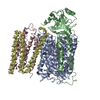
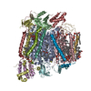
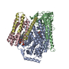
Journal: Nat Commun / Year: 2022 Title: Identifying antibiotics based on structural differences in the conserved allostery from mitochondrial heme-copper oxidases. Authors: Yuya Nishida / Sachiko Yanagisawa / Rikuri Morita / Hideki Shigematsu / Kyoko Shinzawa-Itoh / Hitomi Yuki / Satoshi Ogasawara / Ken Shimuta / Takashi Iwamoto / Chisa Nakabayashi / Waka ...Authors: Yuya Nishida / Sachiko Yanagisawa / Rikuri Morita / Hideki Shigematsu / Kyoko Shinzawa-Itoh / Hitomi Yuki / Satoshi Ogasawara / Ken Shimuta / Takashi Iwamoto / Chisa Nakabayashi / Waka Matsumura / Hisakazu Kato / Chai Gopalasingam / Takemasa Nagao / Tasneem Qaqorh / Yusuke Takahashi / Satoru Yamazaki / Katsumasa Kamiya / Ryuhei Harada / Nobuhiro Mizuno / Hideyuki Takahashi / Yukihiro Akeda / Makoto Ohnishi / Yoshikazu Ishii / Takashi Kumasaka / Takeshi Murata / Kazumasa Muramoto / Takehiko Tosha / Yoshitsugu Shiro / Teruki Honma / Yasuteru Shigeta / Minoru Kubo / Seiji Takashima / Yasunori Shintani / Abstract: Antimicrobial resistance (AMR) is a global health problem. Despite the enormous efforts made in the last decade, threats from some species, including drug-resistant Neisseria gonorrhoeae, continue to ...Antimicrobial resistance (AMR) is a global health problem. Despite the enormous efforts made in the last decade, threats from some species, including drug-resistant Neisseria gonorrhoeae, continue to rise and would become untreatable. The development of antibiotics with a different mechanism of action is seriously required. Here, we identified an allosteric inhibitory site buried inside eukaryotic mitochondrial heme-copper oxidases (HCOs), the essential respiratory enzymes for life. The steric conformation around the binding pocket of HCOs is highly conserved among bacteria and eukaryotes, yet the latter has an extra helix. This structural difference in the conserved allostery enabled us to rationally identify bacterial HCO-specific inhibitors: an antibiotic compound against ceftriaxone-resistant Neisseria gonorrhoeae. Molecular dynamics combined with resonance Raman spectroscopy and stopped-flow spectroscopy revealed an allosteric obstruction in the substrate accessing channel as a mechanism of inhibition. Our approach opens fresh avenues in modulating protein functions and broadens our options to overcome AMR.
In the structure databanks used in Yorodumi, some data are registered as the other names, "COVID-19 virus" and "2019-nCoV". Here are the details of the virus and the list of structure data.
Jan 31, 2019. EMDB accession codes are about to change! (news from PDBe EMDB page)
EMDB accession codes are about to change! (news from PDBe EMDB page)
The allocation of 4 digits for EMDB accession codes will soon come to an end. Whilst these codes will remain in use, new EMDB accession codes will include an additional digit and will expand incrementally as the available range of codes is exhausted. The current 4-digit format prefixed with “EMD-” (i.e. EMD-XXXX) will advance to a 5-digit format (i.e. EMD-XXXXX), and so on. It is currently estimated that the 4-digit codes will be depleted around Spring 2019, at which point the 5-digit format will come into force.
The EM Navigator/Yorodumi systems omit the EMD- prefix.
Related info.:Q: What is EMD? / ID/Accession-code notation in Yorodumi/EM Navigator
Yorodumi is a browser for structure data from EMDB, PDB, SASBDB, etc.
This page is also the successor to EM Navigator detail page, and also detail information page/front-end page for Omokage search.
The word "yorodu" (or yorozu) is an old Japanese word meaning "ten thousand". "mi" (miru) is to see.
Related info.:EMDB / PDB / SASBDB / Comparison of 3 databanks / Yorodumi Search / Aug 31, 2016. New EM Navigator & Yorodumi / Yorodumi Papers / Jmol/JSmol / Function and homology information / Changes in new EM Navigator and Yorodumi
 Movie
Movie Controller

 Yorodumi
 Open data
 Basic information
 Map data
 Sample
 Function and homology information
 ubiquinol oxidase (H+-transporting) / cytochrome bo3 ubiquinol oxidase activity / aerobic electron transport chain / oxidoreductase activity, acting on diphenols and related substances as donors, oxygen as acceptor /
 Authors
 Japan, 2 items
  Citation
 Structure visualization
 Downloads & links
 emd_33293.png
 http://ftp.pdbj.org/pub/emdb/structures/EMD-33293


 Z
Z Y
Y X
X


 Sample components

 Processing
 Electron microscopy