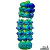
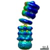
Journal: J Mol Biol / Year: 2020 Title: Cryo-EM Structure of a Bacterial Lipid Transporter YebT. Authors: Chuan Liu / Jinying Ma / Jia Wang / Hongwei Wang / Li Zhang / Abstract: The outer membrane (OM) of Gram-negative bacteria is asymmetric, with lipopolysaccharides (LPSs) on the outer surface and phospholipids (PLs) on the inner surface. This unique organization of OM ...The outer membrane (OM) of Gram-negative bacteria is asymmetric, with lipopolysaccharides (LPSs) on the outer surface and phospholipids (PLs) on the inner surface. This unique organization of OM makes Gram-negative bacteria resistant to many toxic chemicals. How this asymmetric distribution of lipids is maintained has been studied for decades with previous reports of an Mla (Maintenance of OM Lipid Asymmetry) system to be involved. Furthermore, the OM of Gram-negative bacteria is about 20 nm away from inner membrane (IM) where the lipids are synthesized. Therefore, how nascent lipids travel across the periplasmic space and arrive at the inner surface of OM is another interesting question. YebT is a homologue of MlaD in the Mla pathway, but its role in lipid distribution of the OM and IM is largely unknown. Here we report the first high-resolution (~3.0 Å) cryo-EM structure of full-length E. coli YebT in a substrate-bound state. Our structure with details of lipid interaction indicates that YebT is a lipid transporter spanning between IM and OM. We also demonstrate the symmetry mismatch in YebT and the existence of many other conformations of YebT revealing the intrinsic dynamics of this lipid channel. And a brief discussion on possible mechanisms of lipid transport is also included.
History
Deposition
Sep 23, 2019
-
Header (metadata) release
Jan 15, 2020
-
Map release
Jan 15, 2020
-
Update
Mar 11, 2020
-
Current status
Mar 11, 2020
Processing site: PDBj / Status: Released
-
Structure visualization
Movie
Surface view with section colored by density value
In the structure databanks used in Yorodumi, some data are registered as the other names, "COVID-19 virus" and "2019-nCoV". Here are the details of the virus and the list of structure data.
Jan 31, 2019. EMDB accession codes are about to change! (news from PDBe EMDB page)
EMDB accession codes are about to change! (news from PDBe EMDB page)
The allocation of 4 digits for EMDB accession codes will soon come to an end. Whilst these codes will remain in use, new EMDB accession codes will include an additional digit and will expand incrementally as the available range of codes is exhausted. The current 4-digit format prefixed with “EMD-” (i.e. EMD-XXXX) will advance to a 5-digit format (i.e. EMD-XXXXX), and so on. It is currently estimated that the 4-digit codes will be depleted around Spring 2019, at which point the 5-digit format will come into force.
The EM Navigator/Yorodumi systems omit the EMD- prefix.
Related info.:Q: What is EMD? / ID/Accession-code notation in Yorodumi/EM Navigator
Yorodumi is a browser for structure data from EMDB, PDB, SASBDB, etc.
This page is also the successor to EM Navigator detail page, and also detail information page/front-end page for Omokage search.
The word "yorodu" (or yorozu) is an old Japanese word meaning "ten thousand". "mi" (miru) is to see.
Related info.:EMDB / PDB / SASBDB / Comparison of 3 databanks / Yorodumi Search / Aug 31, 2016. New EM Navigator & Yorodumi / Yorodumi Papers / Jmol/JSmol / Function and homology information / Changes in new EM Navigator and Yorodumi
 Movie
Movie Controller


 Open data
 Basic information
 Map data
 Sample
 Escherichia coli K-12 (bacteria)
 Authors
 China, 2 items
  Citation
 Structure visualization
 Movie viewer
 Downloads & links
 emd_0788.png
 http://ftp.pdbj.org/pub/emdb/structures/EMD-0788


 Sample components
 Processing
 Electron microscopy
