 Movie
Movie Controller
[English] 日本語
 Yorodumi
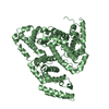
- SASDBK3: Bovine serum albumin, dimer from SEC-SAXS (Bovine serum albumin, ... -
+ Open data

- Basic information
| Entry | Database: SASBDB / ID: SASDBK3 |
|---|---|
 Sample Sample | Bovine serum albumin, dimer from SEC-SAXS
|
| Function / homology |  Function and homology information Function and homology informationcellular response to calcium ion starvation / enterobactin binding / negative regulation of mitochondrial depolarization / toxic substance binding / small molecule binding / cellular response to starvation / fatty acid binding / pyridoxal phosphate binding / blood microparticle / protein-containing complex ...cellular response to calcium ion starvation / enterobactin binding / negative regulation of mitochondrial depolarization / toxic substance binding / small molecule binding / cellular response to starvation / fatty acid binding / pyridoxal phosphate binding / blood microparticle / protein-containing complex / DNA binding / extracellular region / metal ion binding / cytoplasmSimilarity search - Function |
| Biological species |  Bos taurus (cattle) Bos taurus (cattle) |
 Citation Citation | Journal: Nat Protoc / Year: 2016 Title: Preparing monodisperse macromolecular samples for successful biological small-angle X-ray and neutron-scattering experiments. Authors: Cy M Jeffries / Melissa A Graewert / Clément E Blanchet / David B Langley / Andrew E Whitten / Dmitri I Svergun /   Abstract: Small-angle X-ray scattering (SAXS) and small-angle neutron scattering (SANS) are techniques used to extract structural parameters and determine the overall structures and shapes of biological ...Small-angle X-ray scattering (SAXS) and small-angle neutron scattering (SANS) are techniques used to extract structural parameters and determine the overall structures and shapes of biological macromolecules, complexes and assemblies in solution. The scattering intensities measured from a sample contain contributions from all atoms within the illuminated sample volume, including the solvent and buffer components, as well as the macromolecules of interest. To obtain structural information, it is essential to prepare an exactly matched solvent blank so that background scattering contributions can be accurately subtracted from the sample scattering to obtain the net scattering from the macromolecules in the sample. In addition, sample heterogeneity caused by contaminants, aggregates, mismatched solvents, radiation damage or other factors can severely influence and complicate data analysis, so it is essential that the samples be pure and monodisperse for the duration of the experiment. This protocol outlines the basic physics of SAXS and SANS, and it reveals how the underlying conceptual principles of the techniques ultimately 'translate' into practical laboratory guidance for the production of samples of sufficiently high quality for scattering experiments. The procedure describes how to prepare and characterize protein and nucleic acid samples for both SAXS and SANS using gel electrophoresis, size-exclusion chromatography (SEC) and light scattering. Also included are procedures that are specific to X-rays (in-line SEC-SAXS) and neutrons, specifically preparing samples for contrast matching or variation experiments and deuterium labeling of proteins. |
 Contact author Contact author |
|
- Structure visualization
| Structure viewer | Molecule: MolmilJmol/JSmol |
|---|
- Downloads & links
-Data source
| SASBDB page |  SASDBK3 SASDBK3 |
|---|
-Related structure data
| Related structure data | C: citing same article ( |
|---|---|
| Similar structure data |


-External links
| Related items in Molecule of the Month |
|---|


-Models
| Model #448 |   Type: atomic / Radius of dummy atoms: 1.90 A / Chi-square value: 1.044  Search similar-shape structures of this assembly by Omokage search (details) Search similar-shape structures of this assembly by Omokage search (details) |
|---|---|
| Model #450 |   Type: dummy / Radius of dummy atoms: 3.60 A / Chi-square value: 1.104601 / P-value: 0.339000 Search similar-shape structures of this assembly by Omokage search (details) |
-Sample
| Sample | Name: Bovine serum albumin, dimer from SEC-SAXS |
|---|---|
| Buffer | Name: 25 mM Tris 150 mM NaCl 3% (v/v) glycerol / Concentration: 25.00 mM / pH: 7.5 / Composition: 150 mM NaCl 3% v/v glycerol |
| Entity #299 | Name: BSA dimer / Type: protein / Description: Bovine serum albumin, dimer / Formula weight: 66.462 / Num. of mol.: 2 / Source: Bos taurus / References: UniProt: P02769 Sequence: DTHKSEIAHR FKDLGEEHFK GLVLIAFSQY LQQCPFDEHV KLVNELTEFA KTCVADESHA GCEKSLHTLF GDELCKVASL RETYGDMADC CEKQEPERNE CFLSHKDDSP DLPKLKPDPN TLCDEFKADE KKFWGKYLYE IARRHPYFYA PELLYYANKY NGVFQECCQA ...Sequence: DTHKSEIAHR FKDLGEEHFK GLVLIAFSQY LQQCPFDEHV KLVNELTEFA KTCVADESHA GCEKSLHTLF GDELCKVASL RETYGDMADC CEKQEPERNE CFLSHKDDSP DLPKLKPDPN TLCDEFKADE KKFWGKYLYE IARRHPYFYA PELLYYANKY NGVFQECCQA EDKGACLLPK IETMREKVLT SSARQRLRCA SIQKFGERAL KAWSVARLSQ KFPKAEFVEV TKLVTDLTKV HKECCHGDLL ECADDRADLA KYICDNQDTI SSKLKECCDK PLLEKSHCIA EVEKDAIPEN LPPLTADFAE DKDVCKNYQE AKDAFLGSFL YEYSRRHPEY AVSVLLRLAK EYEATLEECC AKDDPHACYS TVFDKLKHLV DEPQNLIKQN CDQFEKLGEY GFQNALIVRY TRKVPQVSTP TLVEVSRSLG KVGTRCCTKP ESERMPCTED YLSLILNRLC VLHEKTPVSE KVTKCCTESL VNRRPCFSAL TPDETYVPKA FDEKLFTFHA DICTLPDTEK QIKKQTALVE LLKHKPKATE EQLKTVMENF VAFVDKCCAA DDKEACFAVE GPKLVVSTQT ALA |
-Experimental information
| Beam | Instrument name: PETRA III EMBL P12 / City: Hamburg / 国: Germany / Type of source: X-ray synchrotronSynchrotron / Wavelength: 0.12 Å / Dist. spec. to detc.: 3.1 mm | |||||||||||||||||||||||||||||||||
|---|---|---|---|---|---|---|---|---|---|---|---|---|---|---|---|---|---|---|---|---|---|---|---|---|---|---|---|---|---|---|---|---|---|---|
| Detector | Name: Pilatus 2M | |||||||||||||||||||||||||||||||||
| Scan | 
| |||||||||||||||||||||||||||||||||
| Distance distribution function P(R) | 
| |||||||||||||||||||||||||||||||||
| Result | 
|
