 Movie
Movie Controller


+ Open data

- Basic information
| Entry |  |
|---|---|
 Sample Sample | MutS tetramer
|
| Function / homology |  Function and homology information Function and homology informationadenine/cytosine mispair binding /  MutS complex / mismatch repair complex / regulation of DNA recombination / mismatched DNA binding / DNA binding, bending / ATP-dependent DNA damage sensor activity / mismatch repair / ADP binding / damaged DNA binding ...adenine/cytosine mispair binding / MutS complex / mismatch repair complex / regulation of DNA recombination / mismatched DNA binding / DNA binding, bending / ATP-dependent DNA damage sensor activity / mismatch repair / ADP binding / damaged DNA binding / DNA damage response / ATP hydrolysis activity / ATP binding / identical protein binding / cytosol MutS complex / mismatch repair complex / regulation of DNA recombination / mismatched DNA binding / DNA binding, bending / ATP-dependent DNA damage sensor activity / mismatch repair / ADP binding / damaged DNA binding ...adenine/cytosine mispair binding / MutS complex / mismatch repair complex / regulation of DNA recombination / mismatched DNA binding / DNA binding, bending / ATP-dependent DNA damage sensor activity / mismatch repair / ADP binding / damaged DNA binding / DNA damage response / ATP hydrolysis activity / ATP binding / identical protein binding / cytosolSimilarity search - Function |
| Biological species |  Escherichia coli (E. coli) Escherichia coli (E. coli) |
 Citation Citation | Journal: Nucleic Acids Res / Year: 2013 Title: Using stable MutS dimers and tetramers to quantitatively analyze DNA mismatch recognition and sliding clamp formation. Authors: Flora S Groothuizen / Alexander Fish / Maxim V Petoukhov / Annet Reumer / Laura Manelyte / Herrie H K Winterwerp / Martin G Marinus / Joyce H G Lebbink / Dmitri I Svergun / Peter Friedhoff / Titia K Sixma /  Abstract: The process of DNA mismatch repair is initiated when MutS recognizes mismatched DNA bases and starts the repair cascade. The Escherichia coli MutS protein exists in an equilibrium between dimers and ...The process of DNA mismatch repair is initiated when MutS recognizes mismatched DNA bases and starts the repair cascade. The Escherichia coli MutS protein exists in an equilibrium between dimers and tetramers, which has compromised biophysical analysis. To uncouple these states, we have generated stable dimers and tetramers, respectively. These proteins allowed kinetic analysis of DNA recognition and structural analysis of the full-length protein by X-ray crystallography and small angle X-ray scattering. Our structural data reveal that the tetramerization domains are flexible with respect to the body of the protein, resulting in mostly extended structures. Tetrameric MutS has a slow dissociation from DNA, which can be due to occasional bending over and binding DNA in its two binding sites. In contrast, the dimer dissociation is faster, primarily dependent on a combination of the type of mismatch and the flanking sequence. In the presence of ATP, we could distinguish two kinetic groups: DNA sequences where MutS forms sliding clamps and those where sliding clamps are not formed efficiently. Interestingly, this inability to undergo a conformational change rather than mismatch affinity is correlated with mismatch repair. |
 Contact author Contact author |
|
- Structure visualization
- Downloads & links
 SASDAX3


-Models
-Sample
| Sample | Name: MutS tetramer / Sample MW: 381 kDa / Specimen concentration: 4.86 mg/ml |
|---|---|
| Buffer | Name: HEPES / Concentration: 50.00 mM / PK: 7 / pH: 7.5 / Comment: 4-(2-hydroxyethyl)-1-piperazineethanesulfonic acid / Composition: KCl 50.000 mM |
| Entity #40 | Name: MutS tetramer / Type: protein / Description: DNA mismatch repair protein MutS / Formula weight: 95.25 / Num. of mol.: 4 / Source: Escherichia coli / References: UniProt: P23909 Sequence: MSAIENFDAH TPMMQQYLRL KAQHPEILLF YRMGDFYELF YDDAKRASQL LDISLTKRGA SAGEPIPMAG IPYHAVENYL AKLVNQGESV AICEQIGDPA TSKGPVERKV VRIVTPGTIS DEALLQERQD NLLAAIWQDS KGFGYATLDI SSGRFRLSEP ADRETMAAEL ...Sequence: MSAIENFDAH TPMMQQYLRL KAQHPEILLF YRMGDFYELF YDDAKRASQL LDISLTKRGA SAGEPIPMAG IPYHAVENYL AKLVNQGESV AICEQIGDPA TSKGPVERKV VRIVTPGTIS DEALLQERQD NLLAAIWQDS KGFGYATLDI SSGRFRLSEP ADRETMAAEL QRTNPAELLY AEDFAEMSLI EGRRGLRRRP LWEFEIDTAR QQLNLQFGTR DLVGFGVENA PRGLCAAGCL LQYAKDTQRT TLPHIRSITM EREQDSIIMD AATRRNLEIT QNLAGGAENT LASVLDCTVT PMGSRMLKRW LHMPVRDTRV LLERQQTIGA LQDFTAGLQP VLRQVGDLER ILARLALRTA RPRDLARMRH AFQQLPELRA QLETVDSAPV QALREKMGEF AELRDLLERA IIDTPPVLVR DGGVIASGYN EELDEWRALA DGATDYLERL EVRERERTGL DTLKVGFNAV HGYYIQISRG QSHLAPINYM RRQTLKNAER YIIPELKEYE DKVLTSKGKA LALEKQLYEE LFDLLLPHLE ALQQSASALA ELDVLVNLAE RAYTLNYTCP TFIDKPGIRI TEGRHPVVEQ VLNEPFIANP LNLSPQRRML IITGPNMGGK STYMRQTALI ALMAYIGSYV PAQKVEIGPI DRIFTRVGAA DDLASGRSTF MVEMTETANI LHNATEYSLV LMDEIGRGTS TYDGLSLAWA CAENLANKIK ALTLFATHYF ELTQLPEKME GVANVHLDAL EHGDTIAFMH SVQDGAASKS YGLAVAALAG VPKEVIKRAR QKLRELESIS PNAAATQVDG TQMSLLSVPE ETSPAVEALE NLDPDSLTPR QALEWIYRLK SLV |
-Experimental information
| Beam | Instrument name: DORIS III X33  / City: Hamburg / 国: Germany / Type of source: X-ray synchrotronSynchrotron / City: Hamburg / 国: Germany / Type of source: X-ray synchrotronSynchrotron | ||||||||||||
|---|---|---|---|---|---|---|---|---|---|---|---|---|---|
| Detector | Name: Pilatus 2M | ||||||||||||
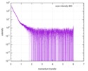
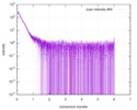
| Scan | 
| ||||||||||||
| Distance distribution function P(R) | 
| ||||||||||||
| Result | 
|
