 Movie
Movie Controller
[English] 日本語
 Yorodumi
- SASDAF5: CYNEX4-T266D (CYNEX4 FRET probe, (eYFP-AnnexinA4-eCFP) T266D mutant) -
+ Open data

- Basic information
| Entry | Database: SASBDB / ID: SASDAF5 |
|---|---|
 Sample Sample | CYNEX4-T266D
|
| Biological species |   Homo sapiens (human) Homo sapiens (human) |
 Citation Citation | Journal: Biophys J / Year: 2012 Title: Conformational analysis of a genetically encoded FRET biosensor by SAXS. Authors: Haydyn D T Mertens / Alen Piljić / Carsten Schultz / Dmitri I Svergun /  Abstract: Genetically encoded FRET (Foerster resonance energy transfer) sensors are exciting tools in modern cell biology. Changes in the conformation of a sensor lead to an altered emission ratio and provide ...Genetically encoded FRET (Foerster resonance energy transfer) sensors are exciting tools in modern cell biology. Changes in the conformation of a sensor lead to an altered emission ratio and provide the means to determine both temporal and spatial changes in target molecules, as well as the activity of enzymes. FRET sensors are widely used to follow phosphorylation events and to monitor the effects of elevated calcium levels. Here, we report for the first time, to our knowledge, on the analysis of the conformational changes involved in sensor function at low resolution using a combination of in vitro and in cellulo FRET measurements and small-angle scattering of x rays (SAXS). The large and dynamic structural rearrangements involved in the modification of the calcium- and phosphorylation-sensitive probe CYNEX4 are comprehensively characterized. It is demonstrated that the synergistic use of SAXS and FRET methods allows one to resolve the ambiguities arising due to the rotation of the sensor molecules and the flexibility of the probe. |
 Contact author Contact author |
|
- Structure visualization
| Structure viewer | Molecule:  MolmilJmol/JSmol MolmilJmol/JSmol |
|---|
- Downloads & links
 SASDAF5
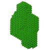


-Models
| Model #112 |   Type: dummy / Software: dammif / Radius of dummy atoms: 2.80 A / Chi-square value: 0.622521  Search similar-shape structures of this assembly by Omokage search (details) Search similar-shape structures of this assembly by Omokage search (details) |
|---|---|
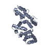
| Model #114 |   Type: mix / Software: coral / Radius of dummy atoms: 1.90 A / Chi-square value: 0.5776 Search similar-shape structures of this assembly by Omokage search (details) |
-Sample
| Sample | Name: CYNEX4-T266D / Sample MW: 92.8 kDa / Specimen concentration: 0.90-17.00 / Concentration method: A280 |
|---|---|
| Buffer | Name: HEPES / Concentration: 50.00 mM / PK: 7 / pH: 7.5 / Comment: 4-(2-hydroxyethyl)-1-piperazineethanesulfonic acid / Composition: KCl 50.000 mM |
| Entity #89 | Name: CYNEX4-T266D / Type: protein Description: CYNEX4 FRET probe, (eYFP-AnnexinA4-eCFP) T266D mutant Formula weight: 92.8 / Num. of mol.: 1 / Source: Homo sapiens Sequence: MASWSHPQFE KGAMASKGEE LFTGVVPILV ELDGDVNGHK FSVSGEGEGD ATYGKLTLKF ICTTGKLPVP WPTLVTTFSY GVQCFSRYPD HMKRHDFFKS AMPEGYVQER TIFFKDDGNY KTRAEVKFEG DTLVNRIELK GIDFKEDGNI LGHKLEYNYN SHNVYIMADK ...Sequence: MASWSHPQFE KGAMASKGEE LFTGVVPILV ELDGDVNGHK FSVSGEGEGD ATYGKLTLKF ICTTGKLPVP WPTLVTTFSY GVQCFSRYPD HMKRHDFFKS AMPEGYVQER TIFFKDDGNY KTRAEVKFEG DTLVNRIELK GIDFKEDGNI LGHKLEYNYN SHNVYIMADK QKNGIKVNFK IRHNIEDGSV QLADHYQQNT PIGDGPVLLP DNHYLSTQSA LSKDPNEKRD HMVLLEFVTA AGITLGMDEL YKRILATMAM ATKGGTVKAA SGFNAMEDAQ TLRKAMKGLG TDEDAIISVL AYRNTAQRQE IRTAYKSTIG RDLIDDLKSE LSGNFEQVIV GMMTPTVLYD VQELRRAMKG AGTDEGCLIE ILASRTPEEI RRISQTYQQQ YGRSLEDDIR SDTSFMFQRV LVSLSAGGRD EGNYLDDALV RQDAQDLYEA GEKKWGTDEV KFLTVLCSRN RNHLLHVFDE YKRISQKDIE QSIKSETSGS FEDALLAIVK CMRNKSAYFA EKLYKSMKGL GTDDNTLIRV MVSRAEIDML DIRAHFKRLY GKSLYSFIKG DTSGDYRKVL LVLCGGDDSR DPPVATMASK GEELFTGVVP ILVELDGDVN GHKFSVSGEG EGDATYGKLT LKFICTTGKL PVPWPTLVTT FSYGVQCFSR YPDHMKRHDF FKSAMPEGYV QERTIFFKDD GNYKTRAEVK FEGDTLVNRI ELKGIDFKED GNILGHKLEY NYNSHNVYIM ADKQKNGIKV NFKIRHNIED GSVQLADHYQ QNTPIGDGPV LLPDNHYLST QSALSKDPNE KRDHMVLLEF VTAAGITLGM DELYK |
-Experimental information
| Beam | Instrument name: DORIS III X33 / City: Hamburg / 国: Germany / Shape: 0.6 / Type of source: X-ray synchrotronSynchrotron / Wavelength: 0.15 Å / Dist. spec. to detc.: 2.7 mm | ||||||||||||||||||||||||||||||
|---|---|---|---|---|---|---|---|---|---|---|---|---|---|---|---|---|---|---|---|---|---|---|---|---|---|---|---|---|---|---|---|
| Detector | Name: Pilatus 1M-W / Pixsize x: 0.172 mm | ||||||||||||||||||||||||||||||
| Scan | 
| ||||||||||||||||||||||||||||||
| Distance distribution function P(R) | 
| ||||||||||||||||||||||||||||||
| Result | 
|
