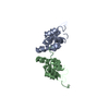
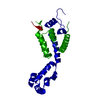
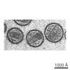
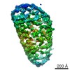
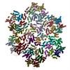
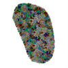
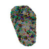
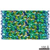
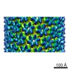
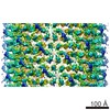
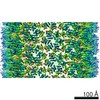
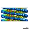
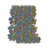
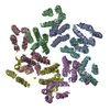
ジャーナル: Nature / 年: 2013 タイトル: Mature HIV-1 capsid structure by cryo-electron microscopy and all-atom molecular dynamics. 著者: Gongpu Zhao / Juan R Perilla / Ernest L Yufenyuy / Xin Meng / Bo Chen / Jiying Ning / Jinwoo Ahn / Angela M Gronenborn / Klaus Schulten / Christopher Aiken / Peijun Zhang / 要旨: Retroviral capsid proteins are conserved structurally but assemble into different morphologies. The mature human immunodeficiency virus-1 (HIV-1) capsid is best described by a 'fullerene cone' model, ...Retroviral capsid proteins are conserved structurally but assemble into different morphologies. The mature human immunodeficiency virus-1 (HIV-1) capsid is best described by a 'fullerene cone' model, in which hexamers of the capsid protein are linked to form a hexagonal surface lattice that is closed by incorporating 12 capsid-protein pentamers. HIV-1 capsid protein contains an amino-terminal domain (NTD) comprising seven α-helices and a β-hairpin, a carboxy-terminal domain (CTD) comprising four α-helices, and a flexible linker with a 310-helix connecting the two structural domains. Structures of the capsid-protein assembly units have been determined by X-ray crystallography; however, structural information regarding the assembled capsid and the contacts between the assembly units is incomplete. Here we report the cryo-electron microscopy structure of a tubular HIV-1 capsid-protein assembly at 8 Å resolution and the three-dimensional structure of a native HIV-1 core by cryo-electron tomography. The structure of the tubular assembly shows, at the three-fold interface, a three-helix bundle with critical hydrophobic interactions. Mutagenesis studies confirm that hydrophobic residues in the centre of the three-helix bundle are crucial for capsid assembly and stability, and for viral infectivity. The cryo-electron-microscopy structures enable modelling by large-scale molecular dynamics simulation, resulting in all-atom models for the hexamer-of-hexamer and pentamer-of-hexamer elements as well as for the entire capsid. Incorporation of pentamers results in closer trimer contacts and induces acute surface curvature. The complete atomic HIV-1 capsid model provides a platform for further studies of capsid function and for targeted pharmacological intervention.
名称: HIV-1 capsid protein / タイプ: COMPLEX / 詳細: hexamer / 別称: HIV CA
分子量
値: 0.025 MDa / 実験値: NO
緩衝液
名称: 1 M NaCl, 50 mM Tris-HCl / pH: 8 / 詳細: 1 M NaCl, 50 mM Tris-HCl
試料
濃度: 2 mg/ml / 包埋: NO / シャドウイング: NO / 染色: NO / 凍結: YES
試料支持
詳細: 200 mesh quantifoil R2/1 copper grid
急速凍結
装置: HOMEMADE PLUNGER / 凍結剤: ETHANE / Temp: 95 K / 湿度: 80 % 詳細: With 2.5 uL sample on carbon side, add 3 uL dilution buffer (100 mM NaCl, 50 mM Tris, pH 8.0) to back side. Blot 3-5 seconds from back side and plunge into liquid ethane with a homemade plunger. 手法: With 2.5 uL sample on carbon side, add 3 uL dilution buffer (100 mM NaCl, 50 mM Tris, pH 8.0) to back side. Blot 3-5 seconds from back side.
モード: BRIGHT FIELDBright-field microscopy / 倍率(公称値): 59000 X / 倍率(補正後): 58257 X / 最大 デフォーカス(公称値): 3500 nm / 最小 デフォーカス(公称値): 1000 nm / Cs: 2 mm / カメラ長: 0 mm
試料ホルダ
試料ホルダーモデル: OTHER / 資料ホルダタイプ: Polara cartridge / 温度: 82 K / 最高温度: 85 K / 最低温度: 80 K / 傾斜角・最大: 0 ° / 傾斜角・最小: 0 °
撮影
電子線照射量: 15 e/Å2 / フィルム・検出器のモデル: KODAK SO-163 FILM
画像スキャン
デジタル画像の数: 27
放射
プロトコル: SINGLE WAVELENGTH / 単色(M)・ラウエ(L): M / 散乱光タイプ: x-ray
放射波長
相対比: 1
-
解析
EMソフトウェア
ID
名称
カテゴリ
1
MDFF
モデルフィッティング
2
FREALIGN
3次元再構成
CTF補正
詳細: each filament
らせん対称
回転角度/サブユニット: 31.13 ° / 軸方向距離/サブユニット: 7.247 Å / らせん対称軸の対称性: C1
3次元再構成
手法: real space helical reconstruction / 解像度: 8.6 Å / 解像度の算出法: FSC 0.5 CUT-OFF / 粒子像の数: 3210 / ピクセルサイズ(実測値): 1.09 Å 詳細: (Helical Details: The segments were aligned and reconstructed using Frealign. Twofold symmetry was imposed using IHRSR++.) 対称性のタイプ: HELICAL
原子モデル構築
ID
プロトコル
空間
詳細
1
FLEXIBLEFIT
REAL
METHOD--Seven hexamers were docked into density then solvated into 1M NaCl. Secondary structure restraints were applied to helices 1 to 11. MDFF was run for 10 ns with symmetry restraints between CA monomers along the chiral axis. The center hexamer was then extracted. PDB entries 3H47 and 2KOD were the starting structures. REFINEMENT PROTOCOL--flexible
2
FLEXIBLEFIT
REAL
METHOD--Seven hexamers were docked into density then solvated into 1M NaCl. Secondary structure restraints were applied to helices 1 to 11. MDFF was run for 10 ns with symmetry restraints between CA monomers along the chiral axis. The center hexamer was then extracted. PDB entries 3H47 and 2KOD were the starting structures. REFINEMENT PROTOCOL--flexible
 ムービー
ムービー コントローラー


 データを開く
 基本情報
基本情報 要素
要素 カプシド
カプシド  キーワード
キーワード 機能・相同性情報
 データ登録者
 引用
引用
 構造の表示
構造の表示 ダウンロードとリンク
 その他のダウンロード
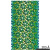
 PDBj
PDBj

 集合体
集合体

 試料調製
試料調製 電子顕微鏡撮影
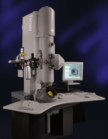
 解析
解析