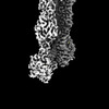
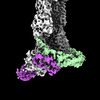
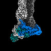
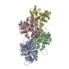
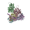
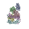
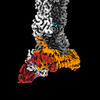
ジャーナル: Science / 年: 2024 タイトル: Molecular mechanism of actin filament elongation by formins. 著者: Wout Oosterheert / Micaela Boiero Sanders / Johanna Funk / Daniel Prumbaum / Stefan Raunser / Peter Bieling / 要旨: Formins control the assembly of actin filaments (F-actin) that drive cell morphogenesis and motility in eukaryotes. However, their molecular interaction with F-actin and their mechanism of action ...Formins control the assembly of actin filaments (F-actin) that drive cell morphogenesis and motility in eukaryotes. However, their molecular interaction with F-actin and their mechanism of action remain unclear. In this work, we present high-resolution cryo-electron microscopy structures of F-actin barbed ends bound by three distinct formins, revealing a common asymmetric formin conformation imposed by the filament. Formation of new intersubunit contacts during actin polymerization sterically displaces formin and triggers its translocation. This "undock-and-lock" mechanism explains how actin-filament growth is coordinated with formin movement. Filament elongation speeds are controlled by the positioning and stability of actin-formin interfaces, which distinguish fast and slow formins. Furthermore, we provide a structure of the actin-formin-profilin ring complex, which resolves how profilin is rapidly released from the barbed end during filament elongation.
タンパク質・ペプチド: Methylated-DNA--protein-cysteine methyltransferase,Cell division control protein 12
複合体: Profilin-1-S29C/S71M
タンパク質・ペプチド: Profilin-1プロフィリン
複合体: Phalloidin
タンパク質・ペプチド: Phalloidin (Amanita phalloides)
リガンド: ADENOSINE-5'-DIPHOSPHATE
リガンド: MAGNESIUM ION
リガンド: PHOSPHATE IONリン酸塩
+
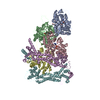
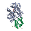
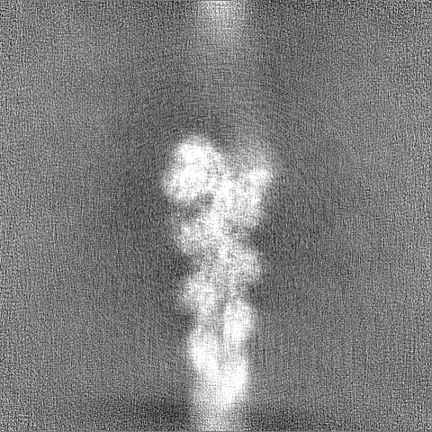
超分子 #1: Actin-formin-profilin ring complex: the phalloidin-stabilized F-a...
超分子
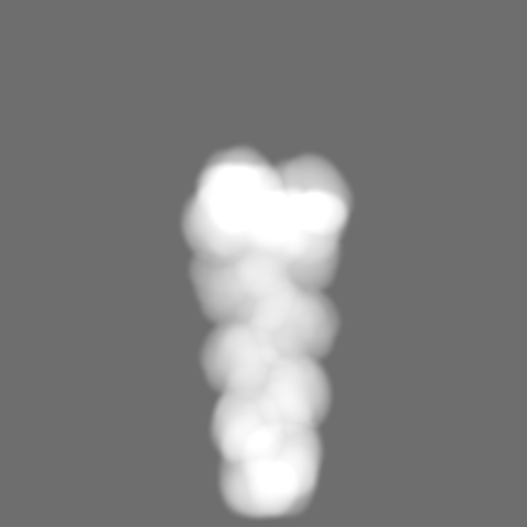
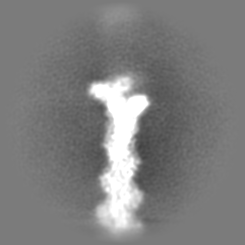
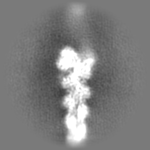
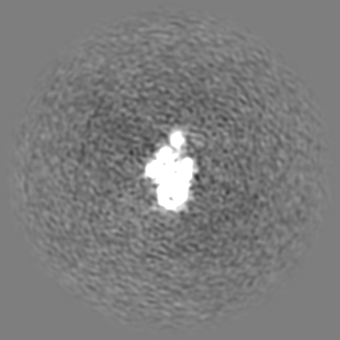
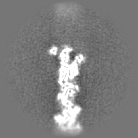
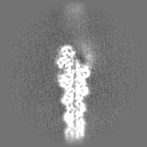
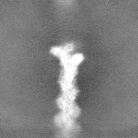
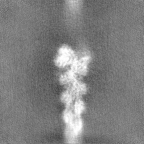
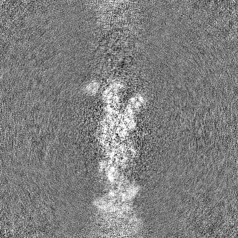
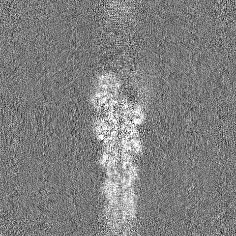
名称: Actin-formin-profilin ring complex: the phalloidin-stabilized F-actin barbed end bound by dimeric-Cdc12 and profilin-S71M. タイプ: complex / ID: 1 / 親要素: 0 / 含まれる分子: #1-#4 詳細: Human beta-actin, human profilin1-S29C/S71M and S. Pombe Cdc12 were purified separately, phalloidin (from Amanita phalloides) was bought from sigma. All components were mixed to assemble the ...詳細: Human beta-actin, human profilin1-S29C/S71M and S. Pombe Cdc12 were purified separately, phalloidin (from Amanita phalloides) was bought from sigma. All components were mixed to assemble the complex prior to cryo-EM grid preparation.
名称: Methylated-DNA--protein-cysteine methyltransferase,Cell division control protein 12 タイプ: protein_or_peptide / ID: 2 詳細: Cdc12(FH1FH2) was purified recombinantly from E. coli with a N-terminal snap-tag. コピー数: 2 / 光学異性体: LEVO EC番号: methylated-DNA-[protein]-cysteine S-methyltransferase
 ムービー
ムービー コントローラー


 データを開く
 基本情報
基本情報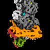
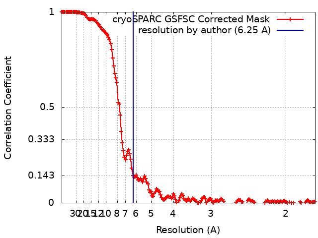
 マップデータ
 試料
試料 キーワード
キーワード actin (アクチン) /
  機能・相同性情報


 データ登録者
 ドイツ, European Union, 3件
  引用
引用 構造の表示
構造の表示
 ダウンロードとリンク
 emd_19499.png
 http://ftp.pdbj.org/pub/emdb/structures/EMD-19499


 Z
Z Y
Y X
X


 試料の構成要素


 解析
解析 電子顕微鏡法