ムービー
ムービー コントローラー


+ データを開く

- 基本情報
基本情報
| 登録情報 | データベース: EMDB / ID: EMD-5193 | |||||||||
|---|---|---|---|---|---|---|---|---|---|---|
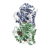
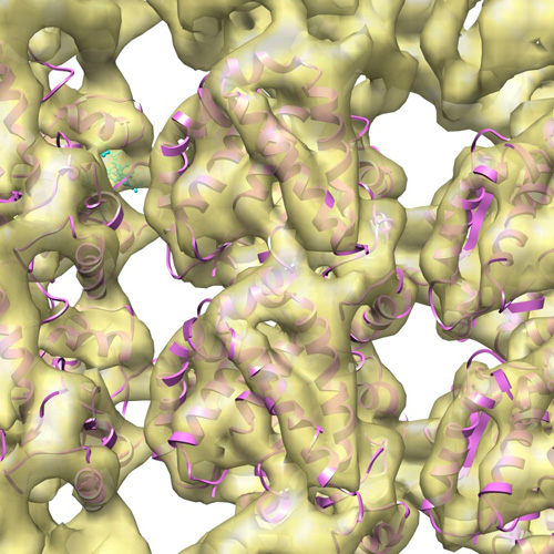
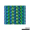
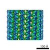
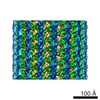
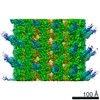
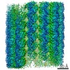
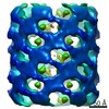
| タイトル | Structural map of MT 13-3 | |||||||||
 マップデータ マップデータ | This is one of a series of six microtubule maps | |||||||||
 試料 試料 |
| |||||||||
 キーワード キーワード | microtubule ultrastructure Cryoelectron Microscopy Computer-Assisted Image Processing | |||||||||
| 生物種 |   Bos taurus (ウシ) Bos taurus (ウシ) | |||||||||
| 手法 | 単粒子再構成法 / クライオ電子顕微鏡法 / 解像度: 9.8 Å | |||||||||
 データ登録者 データ登録者 | Sui H / Downing KH | |||||||||
 引用 引用 | ジャーナル: Structure / 年: 2010 タイトル: Structural basis of interprotofilament interaction and lateral deformation of microtubules. 著者: Haixin Sui / Kenneth H Downing /  要旨: The diverse functions of microtubules require stiff structures possessing sufficient lateral flexibility to enable bending with high curvature. We used cryo-electron microscopy to investigate the ...The diverse functions of microtubules require stiff structures possessing sufficient lateral flexibility to enable bending with high curvature. We used cryo-electron microscopy to investigate the molecular basis for these critical mechanical properties. High-quality structural maps were used to build pseudoatomic models of microtubules containing 11-16 protofilaments, representing a wide range of lateral curvature. Protofilaments in all these microtubules were connected primarily via interprotofilament interactions between the M loops, and the H1'-S2 and H2-S3 loops. We postulate that the tolerance of the loop-loop interactions to lateral deformation provides the capacity for high-curvature bending without breaking. On the other hand, the local molecular architecture that surrounds these connecting loops contributes to the overall rigidity. Interprotofilament interactions in the seam region are similar to those in the normal helical regions, suggesting that the existence of the seam does not significantly affect the mechanical properties of microtubules. | |||||||||
| 履歴 |
|
- 構造の表示
構造の表示
| ムービー |
 ムービービューア ムービービューア |
|---|---|
| 構造ビューア | EMマップ: SurfViewMolmilJmol/JSmol |
| 添付画像 |
- ダウンロードとリンク
-EMDBアーカイブ
| マップデータ | emd_5193.map.gz | 17.3 MB | EMDBマップデータ形式 | |
|---|---|---|---|---|
| ヘッダ (付随情報) | emd-5193-v30.xmlemd-5193.xml | 10.2 KB 10.2 KB | 表示 表示 | EMDBヘッダ |
| 画像 |  emd_5193_1.jpg emd_5193_1.jpg | 153.5 KB | ||
| アーカイブディレクトリ |  http://ftp.pdbj.org/pub/emdb/structures/EMD-5193ftp://ftp.pdbj.org/pub/emdb/structures/EMD-5193 http://ftp.pdbj.org/pub/emdb/structures/EMD-5193ftp://ftp.pdbj.org/pub/emdb/structures/EMD-5193 | HTTPS FTP |
-関連構造データ
-リンク
| EMDBのページ | EMDB (EBI/PDBe) / EMDataResource |
|---|
-マップ
| ファイル | ダウンロード / ファイル: emd_5193.map.gz / 形式: CCP4 / 大きさ: 17.9 MB / タイプ: IMAGE STORED AS FLOATING POINT NUMBER (4 BYTES) | ||||||||||||||||||||||||||||||||||||||||||||||||||||||||||||||||||||
|---|---|---|---|---|---|---|---|---|---|---|---|---|---|---|---|---|---|---|---|---|---|---|---|---|---|---|---|---|---|---|---|---|---|---|---|---|---|---|---|---|---|---|---|---|---|---|---|---|---|---|---|---|---|---|---|---|---|---|---|---|---|---|---|---|---|---|---|---|---|
| 注釈 | This is one of a series of six microtubule maps | ||||||||||||||||||||||||||||||||||||||||||||||||||||||||||||||||||||
| ボクセルのサイズ | X=Y=Z: 2.03 Å | ||||||||||||||||||||||||||||||||||||||||||||||||||||||||||||||||||||
| 密度 |
| ||||||||||||||||||||||||||||||||||||||||||||||||||||||||||||||||||||
| 対称性 | 空間群: 1 | ||||||||||||||||||||||||||||||||||||||||||||||||||||||||||||||||||||
| 詳細 | EMDB XML:
CCP4マップ ヘッダ情報:
| ||||||||||||||||||||||||||||||||||||||||||||||||||||||||||||||||||||
-添付データ
- 試料の構成要素
-全体 : The microtubule containing 13 protofilaments
| 全体 | 名称: The microtubule containing 13 protofilaments |
|---|---|
| 要素 |
|
-超分子 #1000: The microtubule containing 13 protofilaments
| 超分子 | 名称: The microtubule containing 13 protofilaments / タイプ: sample / ID: 1000 集合状態: The 13-protofilament microtubule forms a pseudo helix Number unique components: 2 |
|---|
-分子 #1: microtubule with 13 protofilaments
| 分子 | 名称: microtubule with 13 protofilaments / タイプ: protein_or_peptide / ID: 1 / Name.synonym: MT 13-3 詳細: Microtubules with 13 protofilaments and 3-start helical structure (pseudo-helix) 集合状態: Dimer / 組換発現: No / データベース: NCBI |
|---|---|
| 由来(天然) | 生物種: Bos taurus (ウシ) / 別称: Bovine / 組織: Brain |
| 分子量 | 実験値: 50 MDa / 理論値: 50 MDa |
-実験情報
-構造解析
| 手法 | クライオ電子顕微鏡法 |
|---|---|
 解析 解析 | 単粒子再構成法 |
| 試料の集合状態 | particle |
-試料調製
| 緩衝液 | pH: 6.8 / 詳細: 25mM Pipes, 25mM NaCl, 2mM MgCl2, 1mM EGTA |
|---|---|
| グリッド | 詳細: 300 mesh copper grid covered with home-made carbon films |
| 凍結 | 凍結剤: ETHANE / チャンバー内温度: 93 K / 装置: HOMEMADE PLUNGER / 詳細: Vitrification instrument: Home made plunder / 手法: Blot for 2 seconds before plunging |
- 電子顕微鏡法
| 顕微鏡 | JEOL 4000EX |
|---|---|
| 電子線 | 加速電圧: 400 kV / 電子線源: LAB6 |
| 電子光学系 | 照射モード: FLOOD BEAM / 撮影モード: BRIGHT FIELDBright-field microscopy / Cs: 4.1 mm / 最大 デフォーカス(公称値): 2.0 µm / 最小 デフォーカス(公称値): 0.5 µm / 倍率(公称値): 60000 |
| 試料ステージ | 試料ホルダー: Side entry liquid nitrogen-cooled cryo specimen holder 試料ホルダーモデル: GATAN LIQUID NITROGEN |
| 温度 | 平均: 105 K |
| アライメント法 | Legacy - 非点収差: Objective lens astigmatism was corrected at 400,000 times magnification |
| 日付 | 2007年7月12日 |
| 撮影 | カテゴリ: FILM / フィルム・検出器のモデル: KODAK SO-163 FILM / デジタル化 - スキャナー: OTHER / デジタル化 - サンプリング間隔: 6.35 µm / 実像数: 425 / 平均電子線量: 15 e/Å2 詳細: The micrographs were digitized with a customized robotic scanning system that uses a Nikon Coolpix 8000 scanner ビット/ピクセル: 16 |
-画像解析
| CTF補正 | 詳細: Each micrograph |
|---|---|
| 最終 再構成 | 解像度のタイプ: BY AUTHOR / 解像度: 9.8 Å / 解像度の算出法: OTHER / ソフトウェア - 名称: SPIDER |
| 詳細 | With addition of Taxol or Taxotere for microtubule stabilization |
-原子モデル構築 1
| 初期モデル | PDB ID: |
|---|---|
| ソフトウェア | 名称: Chimera |
| 詳細 | Protocol: Rigid Body. A homology model was generated for the missing region of 1JFF for residuals 35 to 60 in alpha-tubulin (by N. Banavali). The tubulin dimer structure was fitted into the density maps using the rigid body fitting function in Chimera |
| 精密化 | 空間: REAL / プロトコル: RIGID BODY FIT |