 Movie
Movie Controller

[English] 日本語
 Yorodumi
- PDB-9bkz: Crystal Structure of Dephospho-CoA kinase from Klebsiella aerogen... -
+ Open data

- Basic information
| Entry | Database: PDB / ID: 9bkz | |||||||||
|---|---|---|---|---|---|---|---|---|---|---|
| Title | Crystal Structure of Dephospho-CoA kinase from Klebsiella aerogenes (CoA and ADP bound) | |||||||||
 Components Components | Dephospho-CoA kinase | |||||||||
 Keywords Keywords | TRANSFERASE / SSGCID / STRUCTURAL GENOMICS / SEATTLE STRUCTURAL GENOMICS CENTER FOR INFECTIOUS DISEASE | |||||||||
| Function / homology |  Function and homology informationdephospho-CoA kinase / dephospho-CoA kinase activity / coenzyme A biosynthetic process / phosphorylation / ATP binding / cytoplasm Function and homology informationdephospho-CoA kinase / dephospho-CoA kinase activity / coenzyme A biosynthetic process / phosphorylation / ATP binding / cytoplasmSimilarity search - Function | |||||||||
| Biological species |  Klebsiella aerogenes (bacteria) Klebsiella aerogenes (bacteria) | |||||||||
| Method | X-RAY DIFFRACTION / SYNCHROTRON / MOLECULAR REPLACEMENT / Resolution: 2.35 Å | |||||||||
 Authors Authors | Seattle Structural Genomics Center for Infectious Disease / Seattle Structural Genomics Center for Infectious Disease (SSGCID) | |||||||||
| Funding support |  United States, 2items United States, 2items
| |||||||||
 Citation Citation | Journal: To be published Title: Crystal Structure of Dephospho-CoA kinase from Klebsiella aerogenes (CoA and ADP bound) Authors: Liu, L. / Lovell, S. / Battaile, K.P. | |||||||||
| History |
|
- Structure visualization
| Structure viewer | Molecule: MolmilJmol/JSmol |
|---|
- Downloads & links
-Download
| PDBx/mmCIF format | 9bkz.cif.gz | 172.2 KB | Display | PDBx/mmCIF format |
|---|---|---|---|---|
| PDB format | pdb9bkz.ent.gz | 135.5 KB | Display | PDB format |
| PDBx/mmJSON format | 9bkz.json.gz | Tree view | PDBx/mmJSON format | |
| Others |  Other downloads Other downloads |
-Validation report
| Arichive directory | https://data.pdbj.org/pub/pdb/validation_reports/bk/9bkzftp://data.pdbj.org/pub/pdb/validation_reports/bk/9bkz | HTTPS FTP |
|---|
-Related structure data
| Similar structure data |
|---|
-Links
 PDBj
PDBj


- Assembly
| Deposited unit | 
| ||||||||
|---|---|---|---|---|---|---|---|---|---|
| 1 | 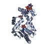
| ||||||||
| 2 | 
| ||||||||
| Unit cell |
|
-Components
| #1: Protein | / Dephosphocoenzyme A kinase Mass: 23850.084 Da / Num. of mol.: 2 / Mutation: G2T, V127I Source method: isolated from a genetically manipulated source Source: (gene. exp.) Klebsiella aerogenes (bacteria) / Gene: coaE, EAE_11320 / Plasmid: KlaeA.00139.a.B1 / Production host: Escherichia coli BL21(DE3) (bacteria) / Strain (production host): BL21(DE3) / References: UniProt: A0A0H3FR62, dephospho-CoA kinase#2: Chemical | Adenosine diphosphate  Mass: 427.201 Da / Num. of mol.: 2 / Source method: obtained synthetically / Formula: C10H15N5O10P2 / Feature type: SUBJECT OF INVESTIGATION / Comment: ADP, energy-carrying molecule*YM Mass: 427.201 Da / Num. of mol.: 2 / Source method: obtained synthetically / Formula: C10H15N5O10P2 / Feature type: SUBJECT OF INVESTIGATION / Comment: ADP, energy-carrying molecule*YM#3: Chemical | ChemComp-COA / | Coenzyme A  Mass: 767.534 Da / Num. of mol.: 1 / Source method: obtained synthetically / Formula: C21H36N7O16P3S / Feature type: SUBJECT OF INVESTIGATION Mass: 767.534 Da / Num. of mol.: 1 / Source method: obtained synthetically / Formula: C21H36N7O16P3S / Feature type: SUBJECT OF INVESTIGATION#4: Chemical | ChemComp-MPD / ( | 2-Methyl-2,4-pentanediol  Mass: 118.174 Da / Num. of mol.: 1 / Source method: obtained synthetically / Formula: C6H14O2 / Comment: precipitant*YM Mass: 118.174 Da / Num. of mol.: 1 / Source method: obtained synthetically / Formula: C6H14O2 / Comment: precipitant*YM#5: Water | ChemComp-HOH / | Water Mass: 18.015 Da / Num. of mol.: 12 / Source method: isolated from a natural source / Formula: H2O Mass: 18.015 Da / Num. of mol.: 12 / Source method: isolated from a natural source / Formula: H2OHas ligand of interest | Y | |
|---|
-Experimental details
-Experiment
| Experiment | Method: X-RAY DIFFRACTION / Number of used crystals: 1 |
|---|
- Sample preparation
| Crystal | Density Matthews: 2.37 Å3/Da / Density % sol: 48.15 % |
|---|---|
| Crystal grow | Temperature: 291 K / Method: vapor diffusion, sitting drop / pH: 6.5 Details: Morpheus F4: 12.5% v/v MPD; 12.5% PEG 1000; 12.5% w/v PEG 3350, 100 mM midazole/MES monohydrate (acid), pH 6.5, 20 mM D-Glucose; 20 mM D-Mannose; 20 mMD-Galactose; 20 mM L-Fucose; 20 mM D- ...Details: Morpheus F4: 12.5% v/v MPD; 12.5% PEG 1000; 12.5% w/v PEG 3350, 100 mM midazole/MES monohydrate (acid), pH 6.5, 20 mM D-Glucose; 20 mM D-Mannose; 20 mMD-Galactose; 20 mM L-Fucose; 20 mM D-Xylose; 20 mM N-Acetyl-D-Glucosamine. KlaeA.00139.a.B1.PW39166 at 24.8 mg/mL. Soak with 5mM ADP and 5 mM CoA in 18.75% v/v MPD; 18.75% PEG 1000; 18.75% w/v PEG 3350 for 10 days. Plate: Liu-S-118 Well G12 , Puck: PSL-0715, Cryo: 18.75% v/v MPD; 18.75% PEG 1000; 18.75% w/v PEG 3350 |
-Data collection
| Diffraction | Mean temperature: 100 K / Serial crystal experiment: N |
|---|---|
| Diffraction source | Source: SYNCHROTRON / Site: NSLS-II / Beamline: 19-ID / Wavelength: 0.9786 Å |
| Detector | Type: DECTRIS EIGER2 XE 9M / Detector: PIXEL / Date: Apr 13, 2024 |
| Radiation | Monochromator: Double Crystal Si 111 / Protocol: SINGLE WAVELENGTH / Monochromatic (M) / Laue (L): M / Scattering type: x-ray |
| Radiation wavelength | Wavelength: 0.9786 Å / Relative weight: 1 |
| Reflection | Resolution: 2.35→39.64 Å / Num. obs: 18892 / % possible obs: 99.9 % / Redundancy: 6.9 % / CC1/2: 0.999 / Rmerge(I) obs: 0.088 / Rpim(I) all: 0.037 / Rrim(I) all: 0.096 / Χ2: 1.03 / Net I/σ(I): 11.8 / Num. measured all: 129858 |
| Reflection shell | Resolution: 2.35→2.43 Å / % possible obs: 100 % / Redundancy: 6.9 % / Rmerge(I) obs: 1.154 / Num. measured all: 12786 / Num. unique obs: 1842 / CC1/2: 0.787 / Rpim(I) all: 0.471 / Rrim(I) all: 1.248 / Χ2: 1.02 / Net I/σ(I) obs: 1.6 |
- Processing
| Software |
| ||||||||||||||||||||||||||||||||||||||||||||||||||||||||||||||||||||||||||||||||||||||||||||||||||||||||||||||||||||||||||||||||||||||||||||||||||||||||||||||||||||||||||||||||||||||||||||||||||||||||
|---|---|---|---|---|---|---|---|---|---|---|---|---|---|---|---|---|---|---|---|---|---|---|---|---|---|---|---|---|---|---|---|---|---|---|---|---|---|---|---|---|---|---|---|---|---|---|---|---|---|---|---|---|---|---|---|---|---|---|---|---|---|---|---|---|---|---|---|---|---|---|---|---|---|---|---|---|---|---|---|---|---|---|---|---|---|---|---|---|---|---|---|---|---|---|---|---|---|---|---|---|---|---|---|---|---|---|---|---|---|---|---|---|---|---|---|---|---|---|---|---|---|---|---|---|---|---|---|---|---|---|---|---|---|---|---|---|---|---|---|---|---|---|---|---|---|---|---|---|---|---|---|---|---|---|---|---|---|---|---|---|---|---|---|---|---|---|---|---|---|---|---|---|---|---|---|---|---|---|---|---|---|---|---|---|---|---|---|---|---|---|---|---|---|---|---|---|---|---|---|---|---|
| Refinement | Method to determine structure: MOLECULAR REPLACEMENT / Resolution: 2.35→39.64 Å / SU ML: 0.34 / Cross valid method: FREE R-VALUE / σ(F): 1.35 / Phase error: 35.77 / Stereochemistry target values: ML
| ||||||||||||||||||||||||||||||||||||||||||||||||||||||||||||||||||||||||||||||||||||||||||||||||||||||||||||||||||||||||||||||||||||||||||||||||||||||||||||||||||||||||||||||||||||||||||||||||||||||||
| Solvent computation | Shrinkage radii: 0.9 Å / VDW probe radii: 1.1 Å / Solvent model: FLAT BULK SOLVENT MODEL | ||||||||||||||||||||||||||||||||||||||||||||||||||||||||||||||||||||||||||||||||||||||||||||||||||||||||||||||||||||||||||||||||||||||||||||||||||||||||||||||||||||||||||||||||||||||||||||||||||||||||
| Refinement step | Cycle: LAST / Resolution: 2.35→39.64 Å
| ||||||||||||||||||||||||||||||||||||||||||||||||||||||||||||||||||||||||||||||||||||||||||||||||||||||||||||||||||||||||||||||||||||||||||||||||||||||||||||||||||||||||||||||||||||||||||||||||||||||||
| Refine LS restraints |
| ||||||||||||||||||||||||||||||||||||||||||||||||||||||||||||||||||||||||||||||||||||||||||||||||||||||||||||||||||||||||||||||||||||||||||||||||||||||||||||||||||||||||||||||||||||||||||||||||||||||||
| LS refinement shell |
| ||||||||||||||||||||||||||||||||||||||||||||||||||||||||||||||||||||||||||||||||||||||||||||||||||||||||||||||||||||||||||||||||||||||||||||||||||||||||||||||||||||||||||||||||||||||||||||||||||||||||
| Refinement TLS params. | Method: refined / Refine-ID: X-RAY DIFFRACTION
| ||||||||||||||||||||||||||||||||||||||||||||||||||||||||||||||||||||||||||||||||||||||||||||||||||||||||||||||||||||||||||||||||||||||||||||||||||||||||||||||||||||||||||||||||||||||||||||||||||||||||
| Refinement TLS group |
|
