 Movie
Movie Controller


+ Open data

- Basic information
| Entry | Database: PDB / ID: 8yy8 | |||||||||
|---|---|---|---|---|---|---|---|---|---|---|
| Title | Fzd7 -Gs complex | |||||||||
 Components Components |
| |||||||||
 Keywords Keywords |  MEMBRANE PROTEIN / GPCR / Class F / Frizzled / Fzd7 / Frizzled 7 / Fzd7-Gs complex MEMBRANE PROTEIN / GPCR / Class F / Frizzled / Fzd7 / Frizzled 7 / Fzd7-Gs complex | |||||||||
| Function / homology |  Function and homology information Function and homology informationnegative regulation of ectodermal cell fate specification / negative regulation of cardiac muscle cell differentiation / mesenchymal to epithelial transition / skeletal muscle satellite cell maintenance involved in skeletal muscle regeneration / Wnt receptor activity / somatic stem cell division / non-canonical Wnt signaling pathway / Wnt-protein binding / positive regulation of epithelial cell proliferation involved in wound healing / WNT5:FZD7-mediated leishmania damping ...negative regulation of ectodermal cell fate specification / negative regulation of cardiac muscle cell differentiation / mesenchymal to epithelial transition / skeletal muscle satellite cell maintenance involved in skeletal muscle regeneration / Wnt receptor activity / somatic stem cell division / non-canonical Wnt signaling pathway / Wnt-protein binding / positive regulation of epithelial cell proliferation involved in wound healing / WNT5:FZD7-mediated leishmania damping / frizzled binding / PCP/CE pathway / regulation of canonical Wnt signaling pathway / Class B/2 (Secretin family receptors) / negative regulation of cell-substrate adhesion / Wnt signaling pathway, planar cell polarity pathway / stem cell population maintenance / canonical Wnt signaling pathway / cellular response to retinoic acid / positive regulation of phosphorylation / phosphatidylinositol-4,5-bisphosphate binding / substrate adhesion-dependent cell spreading / Asymmetric localization of PCP proteins / PDZ domain binding / G protein-coupled receptor activity / positive regulation of JNK cascade / Olfactory Signaling Pathway / Activation of the phototransduction cascade / G beta:gamma signalling through PLC beta / Presynaptic function of Kainate receptors / Thromboxane signalling through TP receptor / G-protein activation / G protein-coupled acetylcholine receptor signaling pathway / Activation of G protein gated Potassium channels / Inhibition of voltage gated Ca2+ channels via Gbeta/gamma subunits / Prostacyclin signalling through prostacyclin receptor / Glucagon signaling in metabolic regulation / G beta:gamma signalling through CDC42 / ADP signalling through P2Y purinoceptor 12 / G beta:gamma signalling through BTK / neuron differentiation / Synthesis, secretion, and inactivation of Glucagon-like Peptide-1 (GLP-1) / Sensory perception of sweet, bitter, and umami (glutamate) taste / photoreceptor disc membrane / Adrenaline,noradrenaline inhibits insulin secretion / Glucagon-type ligand receptors / Vasopressin regulates renal water homeostasis via Aquaporins / G alpha (z) signalling events / cellular response to catecholamine stimulus / Glucagon-like Peptide-1 (GLP1) regulates insulin secretion / ADORA2B mediated anti-inflammatory cytokines production / adenylate cyclase-activating dopamine receptor signaling pathway / ADP signalling through P2Y purinoceptor 1 / G beta:gamma signalling through PI3Kgamma / cellular response to prostaglandin E stimulus / Cooperation of PDCL (PhLP1) and TRiC/CCT in G-protein beta folding / sensory perception of taste / recycling endosome membrane / GPER1 signaling / G-protein beta-subunit binding / Inactivation, recovery and regulation of the phototransduction cascade / heterotrimeric G-protein complex / G alpha (12/13) signalling events / extracellular vesicle / signaling receptor complex adaptor activity / Thrombin signalling through proteinase activated receptors (PARs) / retina development in camera-type eye / GTPase binding / Ca2+ pathway / T cell differentiation in thymus / phospholipase C-activating G protein-coupled receptor signaling pathway / G alpha (i) signalling events / fibroblast proliferation / G alpha (s) signalling events / G alpha (q) signalling events / Ras protein signal transduction / cell population proliferation / Extra-nuclear estrogen signaling / positive regulation of MAPK cascade / G protein-coupled receptor signaling pathway / lysosomal membrane / intracellular membrane-bounded organelle / GTPase activity / synapse / protein-containing complex binding / regulation of DNA-templated transcription / positive regulation of DNA-templated transcription / signal transduction / extracellular exosome / membrane / plasma membrane / cytosol / cytoplasmSimilarity search - Function | |||||||||
| Biological species |  Homo sapiens (human) Homo sapiens (human) | |||||||||
| Method | ELECTRON MICROSCOPY / single particle reconstruction / cryo EM / Resolution: 3.22 Å | |||||||||
 Authors Authors | Chen, B. / Xu, L. / Han, G.W. / Xu, F. | |||||||||
| Funding support |  China, 1items China, 1items
| |||||||||
 Citation Citation | Journal: Cell Res / Year: 2021 Title: Cryo-EM structure of constitutively active human Frizzled 7 in complex with heterotrimeric G. Authors: Lu Xu / Bo Chen / Hannes Schihada / Shane C Wright / Ainoleena Turku / Yiran Wu / Gye-Won Han / Maria Kowalski-Jahn / Pawel Kozielewicz / Carl-Fredrik Bowin / Xianjun Zhang / Chao Li / ...Authors: Lu Xu / Bo Chen / Hannes Schihada / Shane C Wright / Ainoleena Turku / Yiran Wu / Gye-Won Han / Maria Kowalski-Jahn / Pawel Kozielewicz / Carl-Fredrik Bowin / Xianjun Zhang / Chao Li / Michel Bouvier / Gunnar Schulte / Fei Xu /     | |||||||||
| History |
|
- Structure visualization
| Structure viewer | Molecule: MolmilJmol/JSmol |
|---|
- Downloads & links
-Download
| PDBx/mmCIF format | 8yy8.cif.gz | 233.9 KB | Display | PDBx/mmCIF format |
|---|---|---|---|---|
| PDB format | pdb8yy8.ent.gz | 179.9 KB | Display | PDB format |
| PDBx/mmJSON format | 8yy8.json.gz | Tree view | PDBx/mmJSON format | |
| Others |  Other downloads Other downloads |
-Validation report
| Arichive directory | https://data.pdbj.org/pub/pdb/validation_reports/yy/8yy8ftp://data.pdbj.org/pub/pdb/validation_reports/yy/8yy8 | HTTPS FTP |
|---|
-Related structure data
| Related structure data |  31340MC M: map data used to model this data C: citing same article ( |
|---|---|
| Similar structure data |
-Links
 PDBj
PDBj


- Assembly
| Deposited unit | 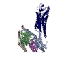
|
|---|---|
| 1 |
|
-Components
| #1: Protein | Mass: 28907.684 Da / Num. of mol.: 1 Source method: isolated from a genetically manipulated source Source: (gene. exp.) Homo sapiens (human) / Production host:  Escherichia coli (E. coli) Escherichia coli (E. coli) |
|---|---|
| #2: Protein | Mass: 37416.930 Da / Num. of mol.: 1 Source method: isolated from a genetically manipulated source Source: (gene. exp.) Homo sapiens (human) / Gene: GNB1 / Production host: Escherichia coli (E. coli) / References: UniProt: P62873 |
| #3: Protein | Mass: 10366.764 Da / Num. of mol.: 1 Source method: isolated from a genetically manipulated source Source: (gene. exp.) Homo sapiens (human) / Gene: GNG2 / Production host: Escherichia coli (E. coli) / References: UniProt: P59768 |
| #4: Protein | Mass: 16054.232 Da / Num. of mol.: 1 Source method: isolated from a genetically manipulated source Source: (gene. exp.) Homo sapiens (human) / Production host: Escherichia coli (E. coli) |
| #5: Protein | / Fz-7 / hFz7 / FzE3 Mass: 66213.539 Da / Num. of mol.: 1 Source method: isolated from a genetically manipulated source Source: (gene. exp.) Homo sapiens (human) / Gene: FZD7 / Production host:   Spodoptera frugiperda (fall armyworm) / References: UniProt: O75084 Spodoptera frugiperda (fall armyworm) / References: UniProt: O75084 |
-Experimental details
-Experiment
| Experiment | Method: ELECTRON MICROSCOPY |
|---|---|
| EM experiment | Aggregation state: PARTICLE / 3D reconstruction method: single particle reconstruction |
- Sample preparation
| Component | Name: human Frizzled 7 in complex with heterotrimeric Gs / Type: COMPLEX / Entity ID: all / Source: RECOMBINANT |
|---|---|
| Molecular weight | Experimental value: NO |
| Source (natural) | Organism: Homo sapiens (human) |
| Source (recombinant) | Organism: Spodoptera frugiperda (fall armyworm) |
| Buffer solution | pH: 7.5 |
| Specimen | Embedding applied: NO / Shadowing applied: NO / Staining applied: NO / Vitrification applied: YES |
| Vitrification | Cryogen name: ETHANE |
- Electron microscopy imaging
| Experimental equipment |  Model: Titan Krios / Image courtesy: FEI Company |
|---|---|
| Microscopy | Model: FEI TITAN KRIOS |
| Electron gun | Electron source: FIELD EMISSION GUN / Accelerating voltage: 300 kV / Illumination mode: FLOOD BEAM |
| Electron lens | Mode: BRIGHT FIELDBright-field microscopy / Nominal magnification: 130000 X / Nominal defocus max: 2200 nm / Nominal defocus min: 700 nm / Cs: 2.7 mm |
| Image recording | Electron dose: 1.5 e/Å2 / Detector mode: SUPER-RESOLUTION / Film or detector model: GATAN K2 SUMMIT (4k x 4k) |
| Image scans | Movie frames/image: 40 |
- Processing
| CTF correction | Type: PHASE FLIPPING AND AMPLITUDE CORRECTION | ||||||||||||||||||||||||
|---|---|---|---|---|---|---|---|---|---|---|---|---|---|---|---|---|---|---|---|---|---|---|---|---|---|
| 3D reconstruction | Resolution: 3.22 Å / Resolution method: FSC 0.143 CUT-OFF / Num. of particles: 224750 / Symmetry type: POINT | ||||||||||||||||||||||||
| Atomic model building | Protocol: AB INITIO MODEL | ||||||||||||||||||||||||
| Refine LS restraints |
|
