 Movie
Movie Controller


+ Open data

- Basic information
| Entry | Database: PDB / ID: 8uxx | ||||||||||||
|---|---|---|---|---|---|---|---|---|---|---|---|---|---|
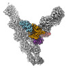
| Title | Arp2/3 branch junction complex, BeFx state | ||||||||||||
 Components Components |
| ||||||||||||
 Keywords Keywords |  CYTOSOLIC PROTEIN / actin / arp2/3 / cytoskeleton / branch CYTOSOLIC PROTEIN / actin / arp2/3 / cytoskeleton / branch | ||||||||||||
| Function / homology |  Function and homology information Function and homology informationRegulation of actin dynamics for phagocytic cup formation / RHO GTPases Activate WASPs and WAVEs / actin cortical patch organization / cell cortex of cell tip / medial cortex / Neutrophil degranulation / establishment or maintenance of cell polarity regulating cell shape / actin cortical patch assembly / Arp2/3 protein complex / Arp2/3 complex-mediated actin nucleation ...Regulation of actin dynamics for phagocytic cup formation / RHO GTPases Activate WASPs and WAVEs / actin cortical patch organization / cell cortex of cell tip / medial cortex / Neutrophil degranulation / establishment or maintenance of cell polarity regulating cell shape / actin cortical patch assembly / Arp2/3 protein complex / Arp2/3 complex-mediated actin nucleation / cell tip / actin cortical patch / regulation of actin filament polymerization / mating projection tip / cortical actin cytoskeleton organization / cytoskeletal motor activator activity / cell division site / tropomyosin binding / myosin heavy chain binding / mesenchyme migration / troponin I binding / actin filament bundle / filamentous actin / actin filament bundle assembly / skeletal muscle thin filament assembly / striated muscle thin filament / mitotic cytokinesis / skeletal muscle myofibril / actin monomer binding / skeletal muscle fiber development / stress fiber / titin binding / actin filament polymerization / filopodium / actin filament / Hydrolases; Acting on acid anhydrides; Acting on acid anhydrides to facilitate cellular and subcellular movement / structural constituent of cytoskeleton / endocytosis / calcium-dependent protein binding / actin filament binding / lamellipodium / cell body / hydrolase activity / protein domain specific binding / calcium ion binding / positive regulation of gene expression / magnesium ion binding / ATP binding / identical protein binding / nucleus / cytosol / cytoplasmSimilarity search - Function | ||||||||||||
| Biological species |  Schizosaccharomyces pombe (fission yeast) Schizosaccharomyces pombe (fission yeast) Oryctolagus cuniculus (rabbit) Oryctolagus cuniculus (rabbit) | ||||||||||||
| Method | ELECTRON MICROSCOPY / single particle reconstruction / cryo EM / Resolution: 3.2 Å | ||||||||||||
 Authors Authors | Chavali, S.S. / Chou, S.Z. / Sindelar, C.V. | ||||||||||||
| Funding support |  United States, 3items United States, 3items
| ||||||||||||
 Citation Citation | Journal: Nat Commun / Year: 2024 Title: Cryo-EM structures reveal how phosphate release from Arp3 weakens actin filament branches formed by Arp2/3 complex. Authors: Sai Shashank Chavali / Steven Z Chou / Wenxiang Cao / Thomas D Pollard / Enrique M De La Cruz / Charles V Sindelar / Abstract: Arp2/3 complex nucleates branched actin filaments for cell and organelle movements. Here we report a 2.7 Å resolution cryo-EM structure of the mature branch junction formed by S. pombe Arp2/3 ...Arp2/3 complex nucleates branched actin filaments for cell and organelle movements. Here we report a 2.7 Å resolution cryo-EM structure of the mature branch junction formed by S. pombe Arp2/3 complex that provides details about interactions with both mother and daughter filaments. We determine a second structure at 3.2 Å resolution with the phosphate analog BeF bound with ADP to Arp3 and ATP bound to Arp2. In this ADP-BeF transition state the outer domain of Arp3 is rotated 2° toward the mother filament compared with the ADP state and makes slightly broader contacts with actin in both the mother and daughter filaments. Thus, dissociation of P from the ADP-P transition state reduces the interactions of Arp2/3 complex with the actin filaments and may contribute to the lower mechanical stability of mature branch junctions with ADP bound to the Arps. Our structures also reveal that the mother filament in contact with Arp2/3 complex is slightly bent and twisted, consistent with the preference of Arp2/3 complex binding curved actin filaments. The small degree of twisting constrains models of actin filament mechanics. | ||||||||||||
| History |
|
- Structure visualization
| Structure viewer | Molecule: MolmilJmol/JSmol |
|---|
- Downloads & links
-Download
| PDBx/mmCIF format | 8uxx.cif.gz | 1.6 MB | Display | PDBx/mmCIF format |
|---|---|---|---|---|
| PDB format | pdb8uxx.ent.gz | 1.3 MB | Display | PDB format |
| PDBx/mmJSON format | 8uxx.json.gz | Tree view | PDBx/mmJSON format | |
| Others |  Other downloads Other downloads |
-Validation report
| Arichive directory | https://data.pdbj.org/pub/pdb/validation_reports/ux/8uxxftp://data.pdbj.org/pub/pdb/validation_reports/ux/8uxx | HTTPS FTP |
|---|
-Related structure data
| Related structure data |  42788MC  8uxwC  8uz0C  8uz1C M: map data used to model this data C: citing same article ( |
|---|---|
| Similar structure data |

-Links
 PDBj
PDBj


- Assembly
| Deposited unit | 
|
|---|---|
| 1 |
|
-Components
-Actin-related protein ... , 7 types, 7 molecules ABCDEFG
| #1: Protein | Mass: 47427.137 Da / Num. of mol.: 1 / Source method: isolated from a natural source / Source: (natural) Schizosaccharomyces pombe (fission yeast) / References: UniProt: P32390 |
|---|---|
| #2: Protein | Mass: 44286.758 Da / Num. of mol.: 1 / Source method: isolated from a natural source / Source: (natural) Schizosaccharomyces pombe (fission yeast) / References: UniProt: Q9UUJ1 |
| #3: Protein | Mass: 41643.465 Da / Num. of mol.: 1 / Source method: isolated from a natural source / Source: (natural) Schizosaccharomyces pombe (fission yeast) / References: UniProt: P78774 |
| #4: Protein | Mass: 37025.230 Da / Num. of mol.: 1 / Source method: isolated from a natural source / Source: (natural) Schizosaccharomyces pombe (fission yeast) / References: UniProt: O14241 |
| #5: Protein | Mass: 19865.746 Da / Num. of mol.: 1 / Source method: isolated from a natural source / Source: (natural) Schizosaccharomyces pombe (fission yeast) / References: UniProt: Q9Y7J4 |
| #6: Protein | Mass: 19637.695 Da / Num. of mol.: 1 / Source method: isolated from a natural source / Source: (natural) Schizosaccharomyces pombe (fission yeast) / References: UniProt: Q92352 |
| #7: Protein | Mass: 16922.059 Da / Num. of mol.: 1 / Source method: isolated from a natural source / Source: (natural) Schizosaccharomyces pombe (fission yeast) / References: UniProt: Q10316 |
-Protein , 1 types, 8 molecules HIMNOPQR
| #8: Protein | / Alpha-actin-1 Mass: 42109.973 Da / Num. of mol.: 8 / Source method: isolated from a natural source / Source: (natural) Oryctolagus cuniculus (rabbit)References: UniProt: P68135, Hydrolases; Acting on acid anhydrides; Acting on acid anhydrides to facilitate cellular and subcellular movement |
|---|
-Non-polymers , 5 types, 32 molecules 


| #9: Chemical | ChemComp-ADP / Adenosine diphosphate Mass: 427.201 Da / Num. of mol.: 9 / Source method: obtained synthetically / Formula: C10H15N5O10P2 / Feature type: SUBJECT OF INVESTIGATION / Comment: ADP, energy-carrying molecule*YM Mass: 427.201 Da / Num. of mol.: 9 / Source method: obtained synthetically / Formula: C10H15N5O10P2 / Feature type: SUBJECT OF INVESTIGATION / Comment: ADP, energy-carrying molecule*YM#10: Chemical | ChemComp-MG /  Mass: 24.305 Da / Num. of mol.: 10 / Source method: obtained synthetically / Formula: Mg / Feature type: SUBJECT OF INVESTIGATION Mass: 24.305 Da / Num. of mol.: 10 / Source method: obtained synthetically / Formula: Mg / Feature type: SUBJECT OF INVESTIGATION#11: Chemical | ChemComp-BEF /  Mass: 66.007 Da / Num. of mol.: 7 / Source method: obtained synthetically / Formula: BeF3 / Feature type: SUBJECT OF INVESTIGATION Mass: 66.007 Da / Num. of mol.: 7 / Source method: obtained synthetically / Formula: BeF3 / Feature type: SUBJECT OF INVESTIGATION#12: Chemical | ChemComp-ATP / | Adenosine triphosphate Mass: 507.181 Da / Num. of mol.: 1 / Source method: obtained synthetically / Formula: C10H16N5O13P3 / Feature type: SUBJECT OF INVESTIGATION / Comment: ATP, energy-carrying molecule*YM Mass: 507.181 Da / Num. of mol.: 1 / Source method: obtained synthetically / Formula: C10H16N5O13P3 / Feature type: SUBJECT OF INVESTIGATION / Comment: ATP, energy-carrying molecule*YM#13: Water | ChemComp-HOH / | WaterMass: 18.015 Da / Num. of mol.: 5 / Source method: isolated from a natural source / Formula: H2O |
|---|
-Details
| Has ligand of interest | Y |
|---|
-Experimental details
-Experiment
| Experiment | Method: ELECTRON MICROSCOPY |
|---|---|
| EM experiment | Aggregation state: PARTICLE / 3D reconstruction method: single particle reconstruction |
- Sample preparation
| Component |
| ||||||||||||||||||||||||||||
|---|---|---|---|---|---|---|---|---|---|---|---|---|---|---|---|---|---|---|---|---|---|---|---|---|---|---|---|---|---|
| Source (natural) |
| ||||||||||||||||||||||||||||
| Buffer solution | pH: 7.5 | ||||||||||||||||||||||||||||
| Specimen | Embedding applied: NO / Shadowing applied: NO / Staining applied: NO / Vitrification applied: YES Details: Actin monomers with bound Ca2+ were converted to Mg2+-actin by equilibrating with 50 micromolar MgCl2 and 0.2 mM EGTA (pH 7.5) for 10 min on ice. Actin was polymerized in the presence of ...Details: Actin monomers with bound Ca2+ were converted to Mg2+-actin by equilibrating with 50 micromolar MgCl2 and 0.2 mM EGTA (pH 7.5) for 10 min on ice. Actin was polymerized in the presence of capping protein (CP) by sequentially mixing 8.75 micromolar Mg-ATP-actin monomers with 0.75 micromolar CP and equilibrated at room temperature for 1 h. In parallel, Arp2/3 complex (0.4 micromolar) in QB buffer was activated by mixing 0.85 micromolar GCN4-VCA and 50 micromolar ATP and incubated at 4 degrees for 1 h. The capped actin filaments sample was then gently mixed with an equal volume of activated Arp2/3 complex sample using cut pipette tips and equilibrated at 4 degrees for 5 min. Daughter filaments were formed and elongated in the presence of CP by adding 0.25 micromolar Mg-actin monomers, 50 micromolar ATP, and 40 nM CP and incubated for 5 min at room temperature. This step was subsequently repeated 4 more times, then aged for ~90 min before preparing grids for cryo-EM data collection. A final concentration of 2 mM BeFx (prepared using 2 mM BeSO4 and 10 mM NaF) was added during the 4th step of daughter filament formation (as mentioned above). | ||||||||||||||||||||||||||||
| Specimen support | Details: Grids were not glow discharged / Grid material: GOLD / Grid mesh size: 300 divisions/in. / Grid type: Quantifoil R1.2/1.3 | ||||||||||||||||||||||||||||
| Vitrification | Instrument: FEI VITROBOT MARK IV / Cryogen name: ETHANE / Humidity: 100 % / Chamber temperature: 277 K Details: The samples were incubated on the grid for 50 s and the extra solution was blotted using two Vitrobot filter papers (0.55/20 mm, Grade 595, Ted Pella) for 4 s at 0 blot force. The grids were ...Details: The samples were incubated on the grid for 50 s and the extra solution was blotted using two Vitrobot filter papers (0.55/20 mm, Grade 595, Ted Pella) for 4 s at 0 blot force. The grids were plunged into liquid ethane at ~180 degrees C with a wait time of 0.5 s. |

- Electron microscopy imaging
| Experimental equipment |  Model: Titan Krios / Image courtesy: FEI Company |
|---|---|
| Microscopy | Model: FEI TITAN KRIOS Details: The vitrified grids were screened for sample homogeneity and ice thickness in a Glacios 200 kV transmission electron microscope equipped with Gatan K2 summit camera. Electron micrographs for ...Details: The vitrified grids were screened for sample homogeneity and ice thickness in a Glacios 200 kV transmission electron microscope equipped with Gatan K2 summit camera. Electron micrographs for image reconstructions were collected using Titan Krios equipped with X-cold field emission gun at 300 kV, Gatan image filter with slit width of 20 eV and a nanoprobe. |
| Electron gun | Electron source: FIELD EMISSION GUN / Accelerating voltage: 300 kV / Illumination mode: FLOOD BEAM |
| Electron lens | Mode: BRIGHT FIELDBright-field microscopy / Nominal magnification: 64000 X / Nominal defocus max: 2500 nm / Nominal defocus min: 1200 nm / Cs: 2.7 mm |
| Specimen holder | Cryogen: NITROGEN / Specimen holder model: FEI TITAN KRIOS AUTOGRID HOLDER |
| Image recording | Average exposure time: 3.3 sec. / Electron dose: 51.4 e/Å2 / Film or detector model: GATAN K3 (6k x 4k) / Num. of real images: 8519 Details: Each movie contains 41 frames with a frame time of 0.08 s. A dose rate of 28.4 counts/pixel/s and a physical pixel size of 1.346 Angstroms was used. |
| EM imaging optics | Energyfilter name: GIF Quantum LS Details: Electron micrographs for image reconstructions were collected using Titan Krios equipped with X-cold field emission gun at 300 kV, Gatan image filter with slit width of 20 eV and a nanoprobe. Energyfilter slit width: 20 eV |
- Processing
| EM software |
| ||||||||||||||||||||
|---|---|---|---|---|---|---|---|---|---|---|---|---|---|---|---|---|---|---|---|---|---|
| CTF correction | Type: PHASE FLIPPING AND AMPLITUDE CORRECTION | ||||||||||||||||||||
| Particle selection | Num. of particles selected: 3000000 | ||||||||||||||||||||
| 3D reconstruction | Resolution: 3.2 Å / Resolution method: FSC 0.143 CUT-OFF / Num. of particles: 200000 / Symmetry type: POINT |
