 Movie
Movie Controller


+ Open data

- Basic information
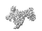
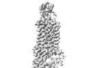
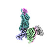
| Entry | Database: PDB / ID: 8tlm | ||||||
|---|---|---|---|---|---|---|---|
| Title | Structure of a class A GPCR/Fab complex | ||||||
 Components Components |
| ||||||
 Keywords Keywords |  STRUCTURAL PROTEIN / GPCR Fab STRUCTURAL PROTEIN / GPCR Fab | ||||||
| Function / homology |  Function and homology informationchemokine receptor activity / C-C chemokine receptor activity / C-C chemokine binding / Chemokine receptors bind chemokines / coreceptor activity / cell chemotaxis / bioluminescence / generation of precursor metabolites and energy / calcium-mediated signaling / chemotaxis ...chemokine receptor activity / C-C chemokine receptor activity / C-C chemokine binding / Chemokine receptors bind chemokines / coreceptor activity / cell chemotaxis / bioluminescence / generation of precursor metabolites and energy / calcium-mediated signaling / chemotaxis / G alpha (i) signalling events / positive regulation of cytosolic calcium ion concentration / cell adhesion / immune response / G protein-coupled receptor signaling pathway / external side of plasma membrane / plasma membrane Function and homology informationchemokine receptor activity / C-C chemokine receptor activity / C-C chemokine binding / Chemokine receptors bind chemokines / coreceptor activity / cell chemotaxis / bioluminescence / generation of precursor metabolites and energy / calcium-mediated signaling / chemotaxis ...chemokine receptor activity / C-C chemokine receptor activity / C-C chemokine binding / Chemokine receptors bind chemokines / coreceptor activity / cell chemotaxis / bioluminescence / generation of precursor metabolites and energy / calcium-mediated signaling / chemotaxis / G alpha (i) signalling events / positive regulation of cytosolic calcium ion concentration / cell adhesion / immune response / G protein-coupled receptor signaling pathway / external side of plasma membrane / plasma membraneSimilarity search - Function | ||||||
| Biological species |  Oryctolagus cuniculus (rabbit) Oryctolagus cuniculus (rabbit) Homo sapiens (human) Homo sapiens (human)  Aequorea victoria (jellyfish) Aequorea victoria (jellyfish) | ||||||
| Method | ELECTRON MICROSCOPY / single particle reconstruction / cryo EM / Resolution: 2.9 Å | ||||||
 Authors Authors | Sun, D. / Johnson, M. / Masureel, M. | ||||||
| Funding support | 1items
| ||||||
 Citation Citation | Journal: Nat Commun / Year: 2023 Title: Structural basis of antibody inhibition and chemokine activation of the human CC chemokine receptor 8. Authors: Dawei Sun / Yonglian Sun / Eric Janezic / Tricia Zhou / Matthew Johnson / Caleigh Azumaya / Sigrid Noreng / Cecilia Chiu / Akiko Seki / Teresita L Arenzana / John M Nicoludis / Yongchang Shi ...Authors: Dawei Sun / Yonglian Sun / Eric Janezic / Tricia Zhou / Matthew Johnson / Caleigh Azumaya / Sigrid Noreng / Cecilia Chiu / Akiko Seki / Teresita L Arenzana / John M Nicoludis / Yongchang Shi / Baomei Wang / Hoangdung Ho / Prajakta Joshi / Christine Tam / Jian Payandeh / Laëtitia Comps-Agrar / Jianyong Wang / Sascha Rutz / James T Koerber / Matthieu Masureel /  Abstract: The C-C motif chemokine receptor 8 (CCR8) is a class A G-protein coupled receptor that has emerged as a promising therapeutic target in cancer. Targeting CCR8 with an antibody has appeared to be an ...The C-C motif chemokine receptor 8 (CCR8) is a class A G-protein coupled receptor that has emerged as a promising therapeutic target in cancer. Targeting CCR8 with an antibody has appeared to be an attractive therapeutic approach, but the molecular basis for chemokine-mediated activation and antibody-mediated inhibition of CCR8 are not fully elucidated. Here, we obtain an antagonist antibody against human CCR8 and determine structures of CCR8 in complex with either the antibody or the endogenous agonist ligand CCL1. Our studies reveal characteristic antibody features allowing recognition of the CCR8 extracellular loops and CCL1-CCR8 interaction modes that are distinct from other chemokine receptor - ligand pairs. Informed by these structural insights, we demonstrate that CCL1 follows a two-step, two-site binding sequence to CCR8 and that antibody-mediated inhibition of CCL1 signaling can occur by preventing the second binding event. Together, our results provide a detailed structural and mechanistic framework of CCR8 activation and inhibition that expands our molecular understanding of chemokine - receptor interactions and offers insight into the development of therapeutic antibodies targeting chemokine GPCRs. | ||||||
| History |
|
- Structure visualization
| Structure viewer | Molecule: MolmilJmol/JSmol |
|---|
- Downloads & links
-Download
| PDBx/mmCIF format | 8tlm.cif.gz | 151.4 KB | Display | PDBx/mmCIF format |
|---|---|---|---|---|
| PDB format | pdb8tlm.ent.gz | 111.1 KB | Display | PDB format |
| PDBx/mmJSON format | 8tlm.json.gz | Tree view | PDBx/mmJSON format | |
| Others |  Other downloads Other downloads |
-Validation report
| Arichive directory | https://data.pdbj.org/pub/pdb/validation_reports/tl/8tlmftp://data.pdbj.org/pub/pdb/validation_reports/tl/8tlm | HTTPS FTP |
|---|
-Related structure data
| Related structure data | 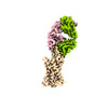 41370MC 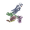 8u1uC M: map data used to model this data C: citing same article ( |
|---|---|
| Similar structure data |
-Links
 PDBj
PDBj


- Assembly
| Deposited unit | 
|
|---|---|
| 1 |
|
-Components
| #1: Antibody | Fragment antigen-binding Mass: 23775.549 Da / Num. of mol.: 1 Source method: isolated from a genetically manipulated source Source: (gene. exp.) Oryctolagus cuniculus (rabbit) / Production host:  Escherichia coli (E. coli) Escherichia coli (E. coli) |
|---|---|
| #2: Antibody | Fragment antigen-binding Mass: 23402.865 Da / Num. of mol.: 1 Source method: isolated from a genetically manipulated source Source: (gene. exp.) Oryctolagus cuniculus (rabbit) / Production host: Escherichia coli (E. coli) |
| #3: Protein | Mass: 76003.508 Da / Num. of mol.: 1 Source method: isolated from a genetically manipulated source Source: (gene. exp.) Homo sapiens (human), (gene. exp.) Aequorea victoria (jellyfish)Gene: CCR8, GFP Production host: Mammalian expression vector Flag-MCS-pcDNA3.1 (others) References: UniProt: P51685, UniProt: P42212 |
-Experimental details
-Experiment
| Experiment | Method: ELECTRON MICROSCOPY |
|---|---|
| EM experiment | Aggregation state: PARTICLE / 3D reconstruction method: single particle reconstruction |
- Sample preparation
| Component | Name: CCR8_Fab complex / Type: COMPLEX / Entity ID: all / Source: MULTIPLE SOURCES |
|---|---|
| Source (natural) | Organism: Homo sapiens (human) |
| Source (recombinant) | Organism: Mammalian expression vector HA-MCS-pcDNA3.1 (others) |
| Buffer solution | pH: 7.5 |
| Specimen | Embedding applied: NO / Shadowing applied: NO / Staining applied: NO / Vitrification applied: YES |
| Vitrification | Cryogen name: ETHANE |
- Electron microscopy imaging
| Experimental equipment |  Model: Titan Krios / Image courtesy: FEI Company |
|---|---|
| Microscopy | Model: FEI TITAN KRIOS |
| Electron gun | Electron source: FIELD EMISSION GUN / Accelerating voltage: 300 kV / Illumination mode: SPOT SCAN |
| Electron lens | Mode: OTHER / Nominal defocus max: 1800 nm / Nominal defocus min: 800 nm |
| Image recording | Electron dose: 1.067 e/Å2 / Film or detector model: FEI FALCON III (4k x 4k) |
- Processing
| CTF correction | Type: PHASE FLIPPING AND AMPLITUDE CORRECTION |
|---|---|
| 3D reconstruction | Resolution: 2.9 Å / Resolution method: FSC 0.143 CUT-OFF / Num. of particles: 340479 / Symmetry type: POINT |
