 Movie
Movie Controller


+ Open data

- Basic information
| Entry | Database: PDB / ID: 8qxj | ||||||||||||
|---|---|---|---|---|---|---|---|---|---|---|---|---|---|
| Title | Cryo-EM structure of tetrameric human SAMHD1 with dApNHpp | ||||||||||||
 Components Components | Deoxynucleoside triphosphate triphosphohydrolase SAMHD1 | ||||||||||||
 Keywords Keywords |  HYDROLASE / TRIPHOSPHOHYDROLASE / METALLO-ENZYME / BINUCLEAR / HD HYDROLASE / TRIPHOSPHOHYDROLASE / METALLO-ENZYME / BINUCLEAR / HD | ||||||||||||
| Function / homology |  Function and homology information Function and homology informationNucleotide catabolism / Hydrolases; Acting on ester bonds; Triphosphoric-monoester hydrolases / deoxynucleoside triphosphate hydrolase activity / dGTP binding / triphosphoric monoester hydrolase activity / dATP catabolic process / dGTPase activity / tetraspanin-enriched microdomain / dGTP catabolic process / DNA strand resection involved in replication fork processing ...Nucleotide catabolism / Hydrolases; Acting on ester bonds; Triphosphoric-monoester hydrolases / deoxynucleoside triphosphate hydrolase activity / dGTP binding / triphosphoric monoester hydrolase activity / dATP catabolic process / dGTPase activity / tetraspanin-enriched microdomain / dGTP catabolic process / DNA strand resection involved in replication fork processing / deoxyribonucleotide catabolic process / negative regulation of type I interferon-mediated signaling pathway / regulation of innate immune response / somatic hypermutation of immunoglobulin genes / RNA nuclease activity / double-strand break repair via homologous recombination / Interferon alpha/beta signaling / single-stranded DNA binding / site of double-strand break / protein homotetramerization / defense response to virus / nucleic acid binding / immune response / innate immune response / DNA damage response / GTP binding / RNA binding / zinc ion binding / nucleoplasm / identical protein binding / nucleus / plasma membraneSimilarity search - Function | ||||||||||||
| Biological species |  Homo sapiens (human) Homo sapiens (human) | ||||||||||||
| Method | ELECTRON MICROSCOPY / single particle reconstruction / cryo EM / Resolution: 2.65 Å | ||||||||||||
 Authors Authors | Acton, O.J. / Sheppard, D. / Rosenthal, P.B. / Taylor, I.A. | ||||||||||||
| Funding support |  United Kingdom, 3items United Kingdom, 3items
| ||||||||||||
 Citation Citation | Journal: Nat Commun / Year: 2024 Title: Platform-directed allostery and quaternary structure dynamics of SAMHD1 catalysis. Authors: Oliver J Acton / Devon Sheppard / Simone Kunzelmann / Sarah J Caswell / Andrea Nans / Ailidh J O Burgess / Geoff Kelly / Elizabeth R Morris / Peter B Rosenthal / Ian A Taylor / Abstract: SAMHD1 regulates cellular nucleotide homeostasis, controlling dNTP levels by catalysing their hydrolysis into 2'-deoxynucleosides and triphosphate. In differentiated CD4+ macrophage and resting T- ...SAMHD1 regulates cellular nucleotide homeostasis, controlling dNTP levels by catalysing their hydrolysis into 2'-deoxynucleosides and triphosphate. In differentiated CD4+ macrophage and resting T-cells SAMHD1 activity results in the inhibition of HIV-1 infection through a dNTP blockade. In cancer, SAMHD1 desensitizes cells to nucleoside-analogue chemotherapies. Here we employ time-resolved cryogenic-EM imaging and single-particle analysis to visualise assembly, allostery and catalysis by this multi-subunit enzyme. Our observations reveal how dynamic conformational changes in the SAMHD1 quaternary structure drive the catalytic cycle. We capture five states at high-resolution in a live catalytic reaction, revealing how allosteric activators support assembly of a stable SAMHD1 tetrameric core and how catalysis is driven by the opening and closing of active sites through pairwise coupling of active sites and order-disorder transitions in regulatory domains. This direct visualisation of enzyme catalysis dynamics within an allostery-stabilised platform sets a precedent for mechanistic studies into the regulation of multi-subunit enzymes. | ||||||||||||
| History |
|
- Structure visualization
| Structure viewer | Molecule: MolmilJmol/JSmol |
|---|
- Downloads & links
-Download
| PDBx/mmCIF format | 8qxj.cif.gz | 358.1 KB | Display | PDBx/mmCIF format |
|---|---|---|---|---|
| PDB format | pdb8qxj.ent.gz | 291.5 KB | Display | PDB format |
| PDBx/mmJSON format | 8qxj.json.gz | Tree view | PDBx/mmJSON format | |
| Others |  Other downloads Other downloads |
-Validation report
| Arichive directory | https://data.pdbj.org/pub/pdb/validation_reports/qx/8qxjftp://data.pdbj.org/pub/pdb/validation_reports/qx/8qxj | HTTPS FTP |
|---|
-Related structure data
| Related structure data |  18729MC 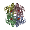 8qxkC 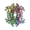 8qxlC  8qxmC 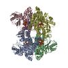 8qxnC  8qxoC M: map data used to model this data C: citing same article ( |
|---|---|
| Similar structure data |


-Links
 PDBj
PDBj

- Assembly
| Deposited unit | 
|
|---|---|
| 1 |
|
-Components
-Protein , 1 types, 4 molecules ABCD
| #1: Protein | Mass: 72305.414 Da / Num. of mol.: 4 Source method: isolated from a genetically manipulated source Source: (gene. exp.) Homo sapiens (human) / Gene: SAMHD1 / Production host:  Escherichia coli (E. coli) / References: UniProt: Q9Y3Z3 Escherichia coli (E. coli) / References: UniProt: Q9Y3Z3 |
|---|
-Non-polymers , 5 types, 242 molecules 


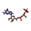

| #2: Chemical | ChemComp-DZ4 /  Mass: 490.197 Da / Num. of mol.: 8 / Source method: obtained synthetically / Formula: C10H17N6O11P3 / Feature type: SUBJECT OF INVESTIGATION Mass: 490.197 Da / Num. of mol.: 8 / Source method: obtained synthetically / Formula: C10H17N6O11P3 / Feature type: SUBJECT OF INVESTIGATION#3: Chemical | ChemComp-FE / Iron Mass: 55.845 Da / Num. of mol.: 4 / Source method: obtained synthetically / Formula: Fe Mass: 55.845 Da / Num. of mol.: 4 / Source method: obtained synthetically / Formula: Fe#4: Chemical | ChemComp-MG /  Mass: 24.305 Da / Num. of mol.: 12 / Source method: obtained synthetically / Formula: Mg Mass: 24.305 Da / Num. of mol.: 12 / Source method: obtained synthetically / Formula: Mg#5: Chemical | ChemComp-GTP / Guanosine triphosphate Mass: 523.180 Da / Num. of mol.: 4 / Source method: obtained synthetically / Formula: C10H16N5O14P3 / Comment: GTP, energy-carrying molecule*YM Mass: 523.180 Da / Num. of mol.: 4 / Source method: obtained synthetically / Formula: C10H16N5O14P3 / Comment: GTP, energy-carrying molecule*YM#6: Water | ChemComp-HOH / | WaterMass: 18.015 Da / Num. of mol.: 214 / Source method: isolated from a natural source / Formula: H2O |
|---|
-Details
| Has ligand of interest | Y |
|---|
-Experimental details
-Experiment
| Experiment | Method: ELECTRON MICROSCOPY |
|---|---|
| EM experiment | Aggregation state: PARTICLE / 3D reconstruction method: single particle reconstruction |
- Sample preparation
| Component | Name: homotetramer of SAMHD1 / Type: COMPLEX / Entity ID: #1 / Source: RECOMBINANT |
|---|---|
| Source (natural) | Organism: Homo sapiens (human) |
| Source (recombinant) | Organism: Escherichia coli (E. coli) |
| Buffer solution | pH: 7.8 |
| Specimen | Embedding applied: NO / Shadowing applied: NO / Staining applied: NO / Vitrification applied: YES |
| Specimen support | Grid material: COPPER / Grid mesh size: 200 divisions/in. / Grid type: Quantifoil R2/2 |
| Vitrification | Instrument: FEI VITROBOT MARK III / Cryogen name: ETHANE / Humidity: 100 % |
- Electron microscopy imaging
| Experimental equipment |  Model: Titan Krios / Image courtesy: FEI Company |
|---|---|
| Microscopy | Model: FEI TITAN KRIOS |
| Electron gun | Electron source: LAB6 / Accelerating voltage: 300 kV / Illumination mode: OTHER |
| Electron lens | Mode: BRIGHT FIELDBright-field microscopy / Nominal magnification: 130000 X / Nominal defocus max: 3500 nm / Nominal defocus min: 1000 nm |
| Image recording | Electron dose: 48.6 e/Å2 / Detector mode: COUNTING / Film or detector model: GATAN K2 SUMMIT (4k x 4k) |
| Image scans | Movie frames/image: 30 |
- Processing
| EM software |
| ||||||||||||||||||||||||
|---|---|---|---|---|---|---|---|---|---|---|---|---|---|---|---|---|---|---|---|---|---|---|---|---|---|
| CTF correction | Type: PHASE FLIPPING AND AMPLITUDE CORRECTION | ||||||||||||||||||||||||
| 3D reconstruction | Resolution: 2.65 Å / Resolution method: FSC 0.143 CUT-OFF / Num. of particles: 139594 / Symmetry type: POINT | ||||||||||||||||||||||||
| Atomic model building | Protocol: RIGID BODY FIT / Space: REAL | ||||||||||||||||||||||||
| Refine LS restraints |
|
