 Movie
Movie Controller


+ Open data

- Basic information
| Entry | Database: PDB / ID: 8qux | ||||||
|---|---|---|---|---|---|---|---|
| Title | Hexameric HIV-1 CA in complex with DDD00100333 | ||||||
 Components Components | Spacer peptide 1 | ||||||
 Keywords Keywords |  VIRAL PROTEIN / Capsid VIRAL PROTEIN / Capsid | ||||||
| Function / homology |  Function and homology information Function and homology informationviral budding via host ESCRT complex / ISG15 antiviral mechanism / host multivesicular body / viral nucleocapsid / host cell nucleus / host cell plasma membrane / virion membrane / structural molecule activity / RNA binding / zinc ion binding / membraneSimilarity search - Function | ||||||
| Biological species |  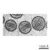 Human immunodeficiency virus 1 Human immunodeficiency virus 1 | ||||||
| Method | X-RAY DIFFRACTION / MOLECULAR REPLACEMENT / Resolution: 2.3 Å | ||||||
 Authors Authors | Petit, A.P. / Fyfe, P.K. | ||||||
| Funding support | 1items
| ||||||
 Citation Citation | Journal: Chemmedchem / Year: 2024 Title: Application of an NMR/crystallography fragment screening platform for the assessment and rapid discovery of new HIV-CA binding fragments. Authors: Lang, S. / Fletcher, D.A. / Petit, A.P. / Luise, N. / Fyfe, P.K. / Zuccotto, F. / Porter, D. / Hope, A. / Bellany, F. / Kerr, C. / MacKenzie, C.J. / Wyatt, P.G. / Gray, D.W. #1: Journal: Biorxiv / Year: 2023Title: Application of an NMR/crystallography fragment screening platform for the assessment and rapid discovery of new HIV-CA binding fragments Authors: Lang, S. / Fletcher, D.A. / Petit, A.P. / Luise, N. / Fyfe, P.K. / Zuccotto, F. / Porter, D. / Hope, D. / Bellany, F. / Kerr, C. / MacKenzie, C.J. / Wyatt, P. / Gray, D.W. | ||||||
| History |
|
- Structure visualization
| Structure viewer | Molecule: MolmilJmol/JSmol |
|---|
- Downloads & links
-Download
| PDBx/mmCIF format | 8qux.cif.gz | 61.2 KB | Display | PDBx/mmCIF format |
|---|---|---|---|---|
| PDB format | pdb8qux.ent.gz | 42.5 KB | Display | PDB format |
| PDBx/mmJSON format | 8qux.json.gz | Tree view | PDBx/mmJSON format | |
| Others |  Other downloads Other downloads |
-Validation report
| Arichive directory | https://data.pdbj.org/pub/pdb/validation_reports/qu/8quxftp://data.pdbj.org/pub/pdb/validation_reports/qu/8qux | HTTPS FTP |
|---|
-Related structure data
| Related structure data | 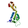 8qubC  8quhC 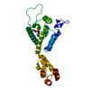 8quiC  8qujC 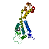 8qukC  8qulC  8quwC  8quyC 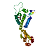 8qv1C  8qv4C  8qv9C  8qvaC C: citing same article ( |
|---|---|
| Similar structure data |

-Links
 PDBj
PDBj

- Assembly
| Deposited unit | 
| ||||||||
|---|---|---|---|---|---|---|---|---|---|
| 1 | x 6
| ||||||||
| Unit cell |
|
-Components
| #1: Protein | Mass: 25461.271 Da / Num. of mol.: 1 Source method: isolated from a genetically manipulated source Source: (gene. exp.) Human immunodeficiency virus 1 / Gene: gag / Production host:  Escherichia coli BL21(DE3) (bacteria) / Variant (production host): C41 / References: UniProt: P12493 Escherichia coli BL21(DE3) (bacteria) / Variant (production host): C41 / References: UniProt: P12493 | ||||||
|---|---|---|---|---|---|---|---|
| #2: Chemical | Ethylene glycol  Mass: 62.068 Da / Num. of mol.: 2 / Source method: obtained synthetically / Formula: C2H6O2 Mass: 62.068 Da / Num. of mol.: 2 / Source method: obtained synthetically / Formula: C2H6O2#3: Chemical | ChemComp-S0I / |   Mass: 238.284 Da / Num. of mol.: 1 / Source method: obtained synthetically / Formula: C15H14N2O / Feature type: SUBJECT OF INVESTIGATION Mass: 238.284 Da / Num. of mol.: 1 / Source method: obtained synthetically / Formula: C15H14N2O / Feature type: SUBJECT OF INVESTIGATION#4: Water | ChemComp-HOH / | Water Mass: 18.015 Da / Num. of mol.: 134 / Source method: isolated from a natural source / Formula: H2O Mass: 18.015 Da / Num. of mol.: 134 / Source method: isolated from a natural source / Formula: H2OHas ligand of interest | Y | |
-Experimental details
-Experiment
| Experiment | Method: X-RAY DIFFRACTION / Number of used crystals: 1 |
|---|
- Sample preparation
| Crystal | Density Matthews: 2.62 Å3/Da / Density % sol: 53.1 % |
|---|---|
| Crystal grow | Temperature: 290 K / Method: vapor diffusion, sitting drop / pH: 8.5 Details: 0.1M Tris buffer, pH 8.0 to 9.0, 10-15% PEG550MME, 0.15M KSCN |
-Data collection
| Diffraction | Mean temperature: 100 K / Serial crystal experiment: N |
|---|---|
| Diffraction source | Source: ROTATING ANODE / Type: RIGAKU MICROMAX-007 HF / Wavelength: 1.54 Å |
| Detector | Type: RIGAKU SATURN 944+ / Detector: CCD / Date: May 2, 2017 |
| Radiation | Protocol: SINGLE WAVELENGTH / Monochromatic (M) / Laue (L): M / Scattering type: x-ray |
| Radiation wavelength | Wavelength: 1.54 Å / Relative weight: 1 |
| Reflection | Resolution: 2.3→28.33 Å / Num. obs: 11849 / % possible obs: 99.9 % / Redundancy: 6.1 % / CC1/2: 0.98 / Rmerge(I) obs: 0.22 / Net I/σ(I): 7.1 |
| Reflection shell | Resolution: 2.3→2.42 Å / Redundancy: 6.1 % / Rmerge(I) obs: 0.84 / Mean I/σ(I) obs: 2.5 / Num. unique obs: 1717 / CC1/2: 0.68 / % possible all: 100 |
- Processing
| Software |
| ||||||||||||||||||||||||||||||||||||||||||||||||||||||||||||||||||||||||||||||||||||||||||||||||||||||||||||||||||||||||||||||||||||||||||||||||||||||||||||||||||||||||||||||||||||||
|---|---|---|---|---|---|---|---|---|---|---|---|---|---|---|---|---|---|---|---|---|---|---|---|---|---|---|---|---|---|---|---|---|---|---|---|---|---|---|---|---|---|---|---|---|---|---|---|---|---|---|---|---|---|---|---|---|---|---|---|---|---|---|---|---|---|---|---|---|---|---|---|---|---|---|---|---|---|---|---|---|---|---|---|---|---|---|---|---|---|---|---|---|---|---|---|---|---|---|---|---|---|---|---|---|---|---|---|---|---|---|---|---|---|---|---|---|---|---|---|---|---|---|---|---|---|---|---|---|---|---|---|---|---|---|---|---|---|---|---|---|---|---|---|---|---|---|---|---|---|---|---|---|---|---|---|---|---|---|---|---|---|---|---|---|---|---|---|---|---|---|---|---|---|---|---|---|---|---|---|---|---|---|---|
| Refinement | Method to determine structure: MOLECULAR REPLACEMENT / Resolution: 2.3→28.33 Å / Cor.coef. Fo:Fc: 0.939 / Cor.coef. Fo:Fc free: 0.878 / SU B: 8.308 / SU ML: 0.195 / Cross valid method: THROUGHOUT / ESU R: 0.326 / ESU R Free: 0.261 / Stereochemistry target values: MAXIMUM LIKELIHOOD / Details: HYDROGENS HAVE BEEN ADDED IN THE RIDING POSITIONS
| ||||||||||||||||||||||||||||||||||||||||||||||||||||||||||||||||||||||||||||||||||||||||||||||||||||||||||||||||||||||||||||||||||||||||||||||||||||||||||||||||||||||||||||||||||||||
| Solvent computation | Ion probe radii: 0.8 Å / Shrinkage radii: 0.8 Å / VDW probe radii: 1.2 Å / Solvent model: MASK | ||||||||||||||||||||||||||||||||||||||||||||||||||||||||||||||||||||||||||||||||||||||||||||||||||||||||||||||||||||||||||||||||||||||||||||||||||||||||||||||||||||||||||||||||||||||
| Displacement parameters | Biso mean: 32.628 Å2
| ||||||||||||||||||||||||||||||||||||||||||||||||||||||||||||||||||||||||||||||||||||||||||||||||||||||||||||||||||||||||||||||||||||||||||||||||||||||||||||||||||||||||||||||||||||||
| Refinement step | Cycle: 1 / Resolution: 2.3→28.33 Å
| ||||||||||||||||||||||||||||||||||||||||||||||||||||||||||||||||||||||||||||||||||||||||||||||||||||||||||||||||||||||||||||||||||||||||||||||||||||||||||||||||||||||||||||||||||||||
| Refine LS restraints |
|