 Movie
Movie Controller

[English] 日本語
 Yorodumi
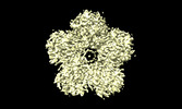
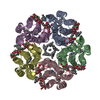
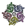
- PDB-8qle: Crystal structure of the light-driven sodium pump ErNaR in the mo... -
+ Open data

- Basic information
| Entry | Database: PDB / ID: 8qle | ||||||
|---|---|---|---|---|---|---|---|
| Title | Crystal structure of the light-driven sodium pump ErNaR in the monomeric form at pH 4.6 | ||||||
 Components Components | Bacteriorhodopsin-like protein | ||||||
 Keywords Keywords |  MEMBRANE PROTEIN / retinal / ion transport / rhodopsin / photocycle / sodium transport MEMBRANE PROTEIN / retinal / ion transport / rhodopsin / photocycle / sodium transport | ||||||
| Function / homology | Bacteriorhodopsin-like protein / Archaeal/bacterial/fungal rhodopsins / Bacteriorhodopsin-like protein / membrane / EICOSANE / OLEIC ACID / Bacteriorhodopsin-like protein Function and homology information Function and homology information | ||||||
| Biological species |  Erythrobacter (bacteria) Erythrobacter (bacteria) | ||||||
| Method | X-RAY DIFFRACTION / SYNCHROTRON / MOLECULAR REPLACEMENT / Resolution: 1.7 Å | ||||||
 Authors Authors | Kovalev, K. / Podoliak, E. / Lamm, G.H.U. / Astashkin, R. / Bourenkov, G. | ||||||
| Funding support |  Germany, 1items Germany, 1items
| ||||||
 Citation Citation | Journal: Nat Commun / Year: 2024 Title: A subgroup of light-driven sodium pumps with an additional Schiff base counterion. Authors: E Podoliak / G H U Lamm / E Marin / A V Schellbach / D A Fedotov / A Stetsenko / M Asido / N Maliar / G Bourenkov / T Balandin / C Baeken / R Astashkin / T R Schneider / A Bateman / J ...Authors: E Podoliak / G H U Lamm / E Marin / A V Schellbach / D A Fedotov / A Stetsenko / M Asido / N Maliar / G Bourenkov / T Balandin / C Baeken / R Astashkin / T R Schneider / A Bateman / J Wachtveitl / I Schapiro / V Busskamp / A Guskov / V Gordeliy / A Alekseev / K Kovalev /     Abstract: Light-driven sodium pumps (NaRs) are unique ion-transporting microbial rhodopsins. The major group of NaRs is characterized by an NDQ motif and has two aspartic acid residues in the central region ...Light-driven sodium pumps (NaRs) are unique ion-transporting microbial rhodopsins. The major group of NaRs is characterized by an NDQ motif and has two aspartic acid residues in the central region essential for sodium transport. Here we identify a subgroup of the NDQ rhodopsins bearing an additional glutamic acid residue in the close vicinity to the retinal Schiff base. We thoroughly characterize a member of this subgroup, namely the protein ErNaR from Erythrobacter sp. HL-111 and show that the additional glutamic acid results in almost complete loss of pH sensitivity for sodium-pumping activity, which is in contrast to previously studied NaRs. ErNaR is capable of transporting sodium efficiently even at acidic pH levels. X-ray crystallography and single particle cryo-electron microscopy reveal that the additional glutamic acid residue mediates the connection between the other two Schiff base counterions and strongly interacts with the aspartic acid of the characteristic NDQ motif. Hence, it reduces its pKa. Our findings shed light on a subgroup of NaRs and might serve as a basis for their rational optimization for optogenetics. | ||||||
| History |
|
- Structure visualization
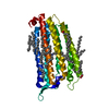
| Structure viewer | Molecule: MolmilJmol/JSmol |
|---|
- Downloads & links
-Download
| PDBx/mmCIF format | 8qle.cif.gz | 76.7 KB | Display | PDBx/mmCIF format |
|---|---|---|---|---|
| PDB format | pdb8qle.ent.gz | 54.5 KB | Display | PDB format |
| PDBx/mmJSON format | 8qle.json.gz | Tree view | PDBx/mmJSON format | |
| Others |  Other downloads Other downloads |
-Validation report
| Arichive directory | https://data.pdbj.org/pub/pdb/validation_reports/ql/8qleftp://data.pdbj.org/pub/pdb/validation_reports/ql/8qle | HTTPS FTP |
|---|
-Related structure data

-Links
 PDBj
PDBj


- Assembly
| Deposited unit | 
| ||||||||
|---|---|---|---|---|---|---|---|---|---|
| 1 |
| ||||||||
| Unit cell |
|
-Components
| #1: Protein | Mass: 32018.041 Da / Num. of mol.: 1 Source method: isolated from a genetically manipulated source Source: (gene. exp.) Erythrobacter (bacteria) / Gene: SAMN04515621_2824 / Production host: Escherichia coli (E. coli) / References: UniProt: A0A1H1XA63 | ||||||
|---|---|---|---|---|---|---|---|
| #2: Chemical | ChemComp-LFA / Icosane  Mass: 282.547 Da / Num. of mol.: 12 / Source method: obtained synthetically / Formula: C20H42 Mass: 282.547 Da / Num. of mol.: 12 / Source method: obtained synthetically / Formula: C20H42#3: Chemical | Oleic acid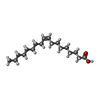  Mass: 282.461 Da / Num. of mol.: 2 / Source method: obtained synthetically / Formula: C18H34O2 Mass: 282.461 Da / Num. of mol.: 2 / Source method: obtained synthetically / Formula: C18H34O2#4: Water | ChemComp-HOH / | Water Mass: 18.015 Da / Num. of mol.: 87 / Source method: isolated from a natural source / Formula: H2O Mass: 18.015 Da / Num. of mol.: 87 / Source method: isolated from a natural source / Formula: H2OHas ligand of interest | Y | |
-Experimental details
-Experiment
| Experiment | Method: X-RAY DIFFRACTION / Number of used crystals: 1 |
|---|
- Sample preparation
| Crystal | Density Matthews: 2.35 Å3/Da / Density % sol: 47.58 % |
|---|---|
| Crystal grow | Temperature: 293 K / Method: lipidic cubic phase / pH: 4.6 / Details: 0.1M sodium acetate pH 4.6, 10% PEG 550MME. |
-Data collection
| Diffraction | Mean temperature: 100 K / Serial crystal experiment: N |
|---|---|
| Diffraction source | Source: SYNCHROTRON / Site: PETRA III, EMBL c/o DESY / Beamline: P14 (MX2) / Wavelength: 0.976 Å |
| Detector | Type: DECTRIS EIGER X 16M / Detector: PIXEL / Date: Mar 29, 2021 |
| Radiation | Protocol: SINGLE WAVELENGTH / Monochromatic (M) / Laue (L): M / Scattering type: x-ray |
| Radiation wavelength | Wavelength: 0.976 Å / Relative weight: 1 |
| Reflection | Resolution: 1.664→60.842 Å / Num. obs: 30115 / % possible obs: 94.2 % / Redundancy: 35.9 % / CC1/2: 0.9987 / Rpim(I) all: 0.052 / Net I/σ(I): 11.038 |
| Reflection shell | Resolution: 1.664→1.763 Å / Mean I/σ(I) obs: 0.698 / Num. unique obs: 1507 / CC1/2: 0.3574 / Rpim(I) all: 1.161 / % possible all: 78.4 |
- Processing
| Software |
| ||||||||||||||||||||||||||||||||||||||||||||||||||||||||||||||||||||||||||||||||||||||||||||||||||||||||||||||||||||||||||||||||||||||||||||||||||||||||||||||||||||||||||||||||||||||
|---|---|---|---|---|---|---|---|---|---|---|---|---|---|---|---|---|---|---|---|---|---|---|---|---|---|---|---|---|---|---|---|---|---|---|---|---|---|---|---|---|---|---|---|---|---|---|---|---|---|---|---|---|---|---|---|---|---|---|---|---|---|---|---|---|---|---|---|---|---|---|---|---|---|---|---|---|---|---|---|---|---|---|---|---|---|---|---|---|---|---|---|---|---|---|---|---|---|---|---|---|---|---|---|---|---|---|---|---|---|---|---|---|---|---|---|---|---|---|---|---|---|---|---|---|---|---|---|---|---|---|---|---|---|---|---|---|---|---|---|---|---|---|---|---|---|---|---|---|---|---|---|---|---|---|---|---|---|---|---|---|---|---|---|---|---|---|---|---|---|---|---|---|---|---|---|---|---|---|---|---|---|---|---|
| Refinement | Method to determine structure: MOLECULAR REPLACEMENT / Resolution: 1.7→20 Å / Cor.coef. Fo:Fc: 0.954 / Cor.coef. Fo:Fc free: 0.943 / SU B: 3.546 / SU ML: 0.108 / Cross valid method: THROUGHOUT / ESU R: 0.141 / ESU R Free: 0.134 / Stereochemistry target values: MAXIMUM LIKELIHOOD / Details: HYDROGENS HAVE BEEN ADDED IN THE RIDING POSITIONS
| ||||||||||||||||||||||||||||||||||||||||||||||||||||||||||||||||||||||||||||||||||||||||||||||||||||||||||||||||||||||||||||||||||||||||||||||||||||||||||||||||||||||||||||||||||||||
| Solvent computation | Ion probe radii: 0.8 Å / Shrinkage radii: 0.8 Å / VDW probe radii: 1.2 Å / Solvent model: MASK | ||||||||||||||||||||||||||||||||||||||||||||||||||||||||||||||||||||||||||||||||||||||||||||||||||||||||||||||||||||||||||||||||||||||||||||||||||||||||||||||||||||||||||||||||||||||
| Displacement parameters | Biso mean: 29.044 Å2
| ||||||||||||||||||||||||||||||||||||||||||||||||||||||||||||||||||||||||||||||||||||||||||||||||||||||||||||||||||||||||||||||||||||||||||||||||||||||||||||||||||||||||||||||||||||||
| Refinement step | Cycle: 1 / Resolution: 1.7→20 Å
| ||||||||||||||||||||||||||||||||||||||||||||||||||||||||||||||||||||||||||||||||||||||||||||||||||||||||||||||||||||||||||||||||||||||||||||||||||||||||||||||||||||||||||||||||||||||
| Refine LS restraints |
|