 Movie
Movie Controller

[English] 日本語
 Yorodumi
- PDB-8q92: P301S Tau Filaments from the Brains of PS19 Transgenic Mouse Line -
+ Open data

- Basic information
| Entry | Database: PDB / ID: 8q92 | |||||||||
|---|---|---|---|---|---|---|---|---|---|---|
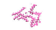
| Title | P301S Tau Filaments from the Brains of PS19 Transgenic Mouse Line | |||||||||
 Components Components | Microtubule-associated protein tau Tau protein Tau protein | |||||||||
 Keywords Keywords | PROTEIN FIBRIL / P301S tau / Frontotemporal dementia and parkinsonism linked to chromosome 17 / Transgenic mice / Electron cryo-microscopy | |||||||||
| Function / homology |  Function and homology information Function and homology informationplus-end-directed organelle transport along microtubule / axonal transport / histone-dependent DNA binding / neurofibrillary tangle assembly / positive regulation of diacylglycerol kinase activity / negative regulation of establishment of protein localization to mitochondrion / neurofibrillary tangle / positive regulation of protein localization to synapse / microtubule lateral binding / tubulin complex ...plus-end-directed organelle transport along microtubule / axonal transport / histone-dependent DNA binding / neurofibrillary tangle assembly / positive regulation of diacylglycerol kinase activity / negative regulation of establishment of protein localization to mitochondrion / neurofibrillary tangle / positive regulation of protein localization to synapse / microtubule lateral binding / tubulin complex / phosphatidylinositol bisphosphate binding / main axon / regulation of long-term synaptic depression / negative regulation of kinase activity / negative regulation of tubulin deacetylation / generation of neurons / regulation of chromosome organization / positive regulation of protein localization / rRNA metabolic process / internal protein amino acid acetylation / regulation of mitochondrial fission / intracellular distribution of mitochondria / axonal transport of mitochondrion / axon development / central nervous system neuron development / regulation of microtubule polymerization / microtubule polymerization / minor groove of adenine-thymine-rich DNA binding / lipoprotein particle binding / dynactin binding / glial cell projection / negative regulation of mitochondrial membrane potential / apolipoprotein binding / protein polymerization / negative regulation of mitochondrial fission / axolemma / Caspase-mediated cleavage of cytoskeletal proteins / regulation of microtubule polymerization or depolymerization / positive regulation of axon extension / Activation of AMPK downstream of NMDARs / regulation of microtubule cytoskeleton organization / supramolecular fiber organization / stress granule assembly / regulation of cellular response to heat / cytoplasmic microtubule organization / axon cytoplasm / regulation of calcium-mediated signaling / positive regulation of microtubule polymerization / somatodendritic compartment / cellular response to brain-derived neurotrophic factor stimulus / synapse assembly / phosphatidylinositol binding / nuclear periphery / cellular response to nerve growth factor stimulus / positive regulation of superoxide anion generation / protein phosphatase 2A binding / regulation of autophagy / astrocyte activation / response to lead ion / synapse organization / microglial cell activation / Hsp90 protein binding / regulation of synaptic plasticity / PKR-mediated signaling / protein homooligomerization / cytoplasmic ribonucleoprotein granule / memory / microtubule cytoskeleton organization / cellular response to reactive oxygen species / SH3 domain binding / activation of cysteine-type endopeptidase activity involved in apoptotic process / neuron projection development / microtubule cytoskeleton / protein-macromolecule adaptor activity / single-stranded DNA binding / cell-cell signaling / cell body / cellular response to heat / actin binding / protein-folding chaperone binding / growth cone / double-stranded DNA binding / microtubule binding / microtubule / sequence-specific DNA binding / amyloid fibril formation / dendritic spine / learning or memory / nuclear speck / neuron projection / membrane raft / axon / negative regulation of gene expression / neuronal cell body / dendrite / DNA damage response / protein kinase binding / enzyme binding / mitochondrion / DNA bindingSimilarity search - Function | |||||||||
| Biological species |  Homo sapiens (human) Homo sapiens (human) | |||||||||
| Method | ELECTRON MICROSCOPY / helical reconstruction / cryo EM / Resolution: 3.05 Å | |||||||||
 Authors Authors | Schweighauser, M. / Murzin, A.G. / Macdonald, J. / Lavenir, I. / Crowther, R.A. / Scheres, S.H.W. / Goedert, M. | |||||||||
| Funding support |  United Kingdom, 2items United Kingdom, 2items
| |||||||||
 Citation Citation | Journal: Acta Neuropathol Commun / Year: 2023 Title: Cryo-EM structures of tau filaments from the brains of mice transgenic for human mutant P301S Tau. Authors: Manuel Schweighauser / Alexey G Murzin / Jennifer Macdonald / Isabelle Lavenir / R Anthony Crowther / Sjors H W Scheres / Michel Goedert / Abstract: Mice transgenic for human mutant P301S tau are widely used as models for human tauopathies. They develop neurodegeneration and abundant filamentous inclusions made of human mutant four-repeat tau. ...Mice transgenic for human mutant P301S tau are widely used as models for human tauopathies. They develop neurodegeneration and abundant filamentous inclusions made of human mutant four-repeat tau. Here we used electron cryo-microscopy (cryo-EM) to determine the structures of tau filaments from the brains of Tg2541 and PS19 mice. Both lines express human P301S tau (0N4R for Tg2541 and 1N4R for PS19) on mixed genetic backgrounds and downstream of different promoters (murine Thy1 for Tg2541 and murine Prnp for PS19). The structures of tau filaments from Tg2541 and PS19 mice differ from each other and those of wild-type tau filaments from human brains. The structures of tau filaments from the brains of humans with mutations P301L, P301S or P301T in MAPT are not known. Filaments from the brains of Tg2541 and PS19 mice share a substructure at the junction of repeats 2 and 3, which comprises residues I297-V312 of tau and includes the P301S mutation. The filament core from the brainstem of Tg2541 mice consists of residues K274-H329 of tau and two disconnected protein densities. Two non-proteinaceous densities are also in evidence. The filament core from the cerebral cortex of line PS19 extends from residues G271-P364 of tau. One strong non-proteinaceous density is also present. Unlike the tau filaments from human brains, the sequences following repeat 4 are missing from the cores of tau filaments from the brains of Tg2541 and PS19 mice. | |||||||||
| History |
|
- Structure visualization
| Structure viewer | Molecule: MolmilJmol/JSmol |
|---|
- Downloads & links
-Download
| PDBx/mmCIF format | 8q92.cif.gz | 63.6 KB | Display | PDBx/mmCIF format |
|---|---|---|---|---|
| PDB format | pdb8q92.ent.gz | 45.7 KB | Display | PDB format |
| PDBx/mmJSON format | 8q92.json.gz | Tree view | PDBx/mmJSON format | |
| Others |  Other downloads Other downloads |
-Validation report
| Arichive directory | https://data.pdbj.org/pub/pdb/validation_reports/q9/8q92ftp://data.pdbj.org/pub/pdb/validation_reports/q9/8q92 | HTTPS FTP |
|---|
-Related structure data
| Related structure data |  18268MC 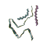 8q96C M: map data used to model this data C: citing same article ( |
|---|---|
| Similar structure data |
-Links
 PDBj
PDBj


- Assembly
| Deposited unit | 
|
|---|---|
| 1 |
|
-Components
| #1: Protein | Tau protein / Neurofibrillary tangle protein / Paired helical filament-tau / PHF-tau Mass: 9967.430 Da / Num. of mol.: 3 / Mutation: P301S Source method: isolated from a genetically manipulated source Source: (gene. exp.) Homo sapiens (human) / Strain: Transgenic PS19 / Tissue: Cerebral Cortex / Gene: MAPT, MAPTL, MTBT1, TAU / Organ: Brain / Production host:  Mus musculus (house mouse) / References: UniProt: P10636 Mus musculus (house mouse) / References: UniProt: P10636 |
|---|
-Experimental details
-Experiment
| Experiment | Method: ELECTRON MICROSCOPY |
|---|---|
| EM experiment | Aggregation state: FILAMENT / 3D reconstruction method: helical reconstruction |
- Sample preparation
| Component | Name: P301S Tau Protein Filament (PS19) / Type: TISSUE Details: Human P301S tau filaments extracted from the brains of transgenic mouse line PS19 Entity ID: all / Source: NATURAL |
|---|---|
| Source (natural) | Organism: Mus musculus (house mouse) / Strain: PS19 / Organ: Brain / Tissue: Cerebral Cortex |
| Buffer solution | pH: 7.4 |
| Specimen | Embedding applied: NO / Shadowing applied: NO / Staining applied: NO / Vitrification applied: YES |
| Specimen support | Grid material: GOLD / Grid mesh size: 300 divisions/in. / Grid type: Quantifoil R1.2/1.3 |
| Vitrification | Instrument: FEI VITROBOT MARK IV / Cryogen name: ETHANE / Humidity: 100 % / Chamber temperature: 277 K |
- Electron microscopy imaging
| Experimental equipment |  Model: Titan Krios / Image courtesy: FEI Company |
|---|---|
| Microscopy | Model: FEI TITAN KRIOS |
| Electron gun | Electron source: FIELD EMISSION GUN / Accelerating voltage: 300 kV / Illumination mode: FLOOD BEAM |
| Electron lens | Mode: BRIGHT FIELDBright-field microscopy / Nominal magnification: 96000 X / Nominal defocus max: 2400 nm / Nominal defocus min: 1500 nm / Alignment procedure: COMA FREE |
| Specimen holder | Cryogen: NITROGEN |
| Image recording | Electron dose: 30 e/Å2 / Film or detector model: FEI FALCON IV (4k x 4k) |
- Processing
| EM software |
| ||||||||||||||||||||||||||||||||||||||||||||||||||||||||||||||||||||||||||||||||||||||||||||||||||||||||||
|---|---|---|---|---|---|---|---|---|---|---|---|---|---|---|---|---|---|---|---|---|---|---|---|---|---|---|---|---|---|---|---|---|---|---|---|---|---|---|---|---|---|---|---|---|---|---|---|---|---|---|---|---|---|---|---|---|---|---|---|---|---|---|---|---|---|---|---|---|---|---|---|---|---|---|---|---|---|---|---|---|---|---|---|---|---|---|---|---|---|---|---|---|---|---|---|---|---|---|---|---|---|---|---|---|---|---|---|
| CTF correction | Type: PHASE FLIPPING AND AMPLITUDE CORRECTION | ||||||||||||||||||||||||||||||||||||||||||||||||||||||||||||||||||||||||||||||||||||||||||||||||||||||||||
| Helical symmerty | Angular rotation/subunit: -0.98 ° / Axial rise/subunit: 4.71 Å / Axial symmetry: C1 | ||||||||||||||||||||||||||||||||||||||||||||||||||||||||||||||||||||||||||||||||||||||||||||||||||||||||||
| 3D reconstruction | Resolution: 3.05 Å / Resolution method: FSC 0.143 CUT-OFF / Num. of particles: 36951 / Symmetry type: HELICAL | ||||||||||||||||||||||||||||||||||||||||||||||||||||||||||||||||||||||||||||||||||||||||||||||||||||||||||
| Refinement | Resolution: 3.05→138.43 Å / Cor.coef. Fo:Fc: 0.761 / SU B: 20.147 / SU ML: 0.342 / ESU R: 0.228 Stereochemistry target values: MAXIMUM LIKELIHOOD WITH PHASES Details: HYDROGENS HAVE BEEN ADDED IN THE RIDING POSITIONS
| ||||||||||||||||||||||||||||||||||||||||||||||||||||||||||||||||||||||||||||||||||||||||||||||||||||||||||
| Solvent computation | Solvent model: PARAMETERS FOR MASK CACLULATION | ||||||||||||||||||||||||||||||||||||||||||||||||||||||||||||||||||||||||||||||||||||||||||||||||||||||||||
| Displacement parameters | Biso mean: 53.879 Å2 | ||||||||||||||||||||||||||||||||||||||||||||||||||||||||||||||||||||||||||||||||||||||||||||||||||||||||||
| Refinement step | Cycle: 1 / Total: 697 | ||||||||||||||||||||||||||||||||||||||||||||||||||||||||||||||||||||||||||||||||||||||||||||||||||||||||||
| Refine LS restraints |
|