 Movie
Movie Controller

[English] 日本語
 Yorodumi
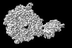
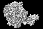
- PDB-8ojb: HSV-1 DNA polymerase-processivity factor complex in exonuclease s... -
+ Open data

- Basic information
| Entry | Database: PDB / ID: 8ojb | |||||||||||||||||||||
|---|---|---|---|---|---|---|---|---|---|---|---|---|---|---|---|---|---|---|---|---|---|---|
| Title | HSV-1 DNA polymerase-processivity factor complex in exonuclease state active site | |||||||||||||||||||||
 Components Components |
| |||||||||||||||||||||
 Keywords Keywords |  TRANSFERASE / DNA / Polymerase / Complex TRANSFERASE / DNA / Polymerase / Complex | |||||||||||||||||||||
| Function / homology |  Function and homology informationDNA polymerase activity / DNA polymerase complex / 5'-3' exonuclease activity / bidirectional double-stranded viral DNA replication / ribonuclease H / RNA-DNA hybrid ribonuclease activity / DNA replication / DNA-directed DNA polymerase / DNA-directed DNA polymerase activity / nucleotide binding ...DNA polymerase activity / DNA polymerase complex / 5'-3' exonuclease activity / bidirectional double-stranded viral DNA replication / ribonuclease H / RNA-DNA hybrid ribonuclease activity / DNA replication / DNA-directed DNA polymerase / DNA-directed DNA polymerase activity / nucleotide binding / host cell nucleus / DNA binding Function and homology informationDNA polymerase activity / DNA polymerase complex / 5'-3' exonuclease activity / bidirectional double-stranded viral DNA replication / ribonuclease H / RNA-DNA hybrid ribonuclease activity / DNA replication / DNA-directed DNA polymerase / DNA-directed DNA polymerase activity / nucleotide binding ...DNA polymerase activity / DNA polymerase complex / 5'-3' exonuclease activity / bidirectional double-stranded viral DNA replication / ribonuclease H / RNA-DNA hybrid ribonuclease activity / DNA replication / DNA-directed DNA polymerase / DNA-directed DNA polymerase activity / nucleotide binding / host cell nucleus / DNA bindingSimilarity search - Function | |||||||||||||||||||||
| Biological species |   Human alphaherpesvirus 1 strain KOS Human alphaherpesvirus 1 strain KOSsynthetic construct (others) | |||||||||||||||||||||
| Method | ELECTRON MICROSCOPY / single particle reconstruction / cryo EM / Resolution: 1.9 Å | |||||||||||||||||||||
 Authors Authors | Gustavsson, E. / Grunewald, K. / Elias, P. / Hallberg, B.M. | |||||||||||||||||||||
| Funding support |  Sweden, Sweden,  Germany, 6items Germany, 6items
| |||||||||||||||||||||
 Citation Citation | Journal: To Be Published Title: Dynamics of the Herpes simplex virus DNA polymerase holoenzyme during DNA synthesis and proof-reading revealed by Cryo-EM Authors: Gustavsson, E. / Grunewald, K. / Elias, P. / Hallberg, B.M. | |||||||||||||||||||||
| History |
|
- Structure visualization
| Structure viewer | Molecule: MolmilJmol/JSmol |
|---|
- Downloads & links
-Download
| PDBx/mmCIF format | 8ojb.cif.gz | 193.9 KB | Display | PDBx/mmCIF format |
|---|---|---|---|---|
| PDB format | pdb8ojb.ent.gz | 142.3 KB | Display | PDB format |
| PDBx/mmJSON format | 8ojb.json.gz | Tree view | PDBx/mmJSON format | |
| Others |  Other downloads Other downloads |
-Validation report
| Arichive directory | https://data.pdbj.org/pub/pdb/validation_reports/oj/8ojbftp://data.pdbj.org/pub/pdb/validation_reports/oj/8ojb | HTTPS FTP |
|---|
-Related structure data
| Related structure data | 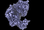 16910MC 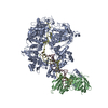 8oj6C  8oj7C 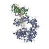 8ojaC  8ojcC  8ojdC  9enpC  9enqC C: citing same article ( M: map data used to model this data |
|---|---|
| Similar structure data |


-Links
 PDBj
PDBj


- Assembly
| Deposited unit | 
|
|---|---|
| 1 |
|
-Components
| #1: DNA chain | Mass: 14260.221 Da / Num. of mol.: 1 / Source method: obtained synthetically / Source: (synth.) synthetic construct (others) | ||||
|---|---|---|---|---|---|
| #2: Protein | Mass: 136683.844 Da / Num. of mol.: 1 Source method: isolated from a genetically manipulated source Source: (gene. exp.) Human alphaherpesvirus 1 strain KOS / Gene: UL30 / Production host:   Spodoptera frugiperda (fall armyworm) Spodoptera frugiperda (fall armyworm)References: UniProt: P04293, DNA-directed DNA polymerase, ribonuclease H | ||||
| #3: Chemical |   Mass: 40.078 Da / Num. of mol.: 2 / Source method: obtained synthetically / Formula: Ca / Feature type: SUBJECT OF INVESTIGATION Mass: 40.078 Da / Num. of mol.: 2 / Source method: obtained synthetically / Formula: Ca / Feature type: SUBJECT OF INVESTIGATION#4: Water | ChemComp-HOH / | Water Mass: 18.015 Da / Num. of mol.: 116 / Source method: isolated from a natural source / Formula: H2O Mass: 18.015 Da / Num. of mol.: 116 / Source method: isolated from a natural source / Formula: H2OHas ligand of interest | Y | |
-Experimental details
-Experiment
| Experiment | Method: ELECTRON MICROSCOPY |
|---|---|
| EM experiment | Aggregation state: PARTICLE / 3D reconstruction method: single particle reconstruction |
- Sample preparation
| Component | Name: HSV-1 DNA polymerase-processivity factor complex in exonuclease state Type: COMPLEX / Entity ID: #2 / Source: RECOMBINANT | |||||||||||||||||||||||||
|---|---|---|---|---|---|---|---|---|---|---|---|---|---|---|---|---|---|---|---|---|---|---|---|---|---|---|
| Molecular weight | Experimental value: NO | |||||||||||||||||||||||||
| Source (natural) | Organism: Human alphaherpesvirus 1 strain KOS | |||||||||||||||||||||||||
| Source (recombinant) | Organism: Spodoptera frugiperda (fall armyworm) | |||||||||||||||||||||||||
| Buffer solution | pH: 7.8 / Details: 20mM HEPES pH7.8, 150mM NaCl, 5mM CaCl2, 2mM DTT | |||||||||||||||||||||||||
| Buffer component |
| |||||||||||||||||||||||||
| Specimen | Conc.: 0.4 mg/ml / Embedding applied: NO / Shadowing applied: NO / Staining applied: NO / Vitrification applied: YES | |||||||||||||||||||||||||
| Specimen support | Grid material: GOLD / Grid mesh size: 300 divisions/in. / Grid type: UltrAuFoil R1.2/1.3 | |||||||||||||||||||||||||
| Vitrification | Instrument: FEI VITROBOT MARK IV / Cryogen name: ETHANE-PROPANE / Humidity: 95 % / Chamber temperature: 277 K |
- Electron microscopy imaging
| Experimental equipment |  Model: Titan Krios / Image courtesy: FEI Company |
|---|---|
| Microscopy | Model: FEI TITAN KRIOS |
| Electron gun | Electron source: FIELD EMISSION GUN / Accelerating voltage: 300 kV / Illumination mode: FLOOD BEAM |
| Electron lens | Mode: BRIGHT FIELDBright-field microscopy / Nominal magnification: 165000 X / Nominal defocus max: 1500 nm / Nominal defocus min: 500 nm / Cs: 2.7 mm / C2 aperture diameter: 70 µm / Alignment procedure: COMA FREE |
| Specimen holder | Cryogen: NITROGEN / Specimen holder model: FEI TITAN KRIOS AUTOGRID HOLDER |
| Image recording | Electron dose: 58 e/Å2 / Film or detector model: GATAN K3 BIOQUANTUM (6k x 4k) |
- Processing
| EM software |
| ||||||||||||||||||||||||||||||||||||||||||||||||||||||||||||||||||||||||||||||||||||||||||||||||||||||||||
|---|---|---|---|---|---|---|---|---|---|---|---|---|---|---|---|---|---|---|---|---|---|---|---|---|---|---|---|---|---|---|---|---|---|---|---|---|---|---|---|---|---|---|---|---|---|---|---|---|---|---|---|---|---|---|---|---|---|---|---|---|---|---|---|---|---|---|---|---|---|---|---|---|---|---|---|---|---|---|---|---|---|---|---|---|---|---|---|---|---|---|---|---|---|---|---|---|---|---|---|---|---|---|---|---|---|---|---|
| CTF correction | Type: PHASE FLIPPING AND AMPLITUDE CORRECTION | ||||||||||||||||||||||||||||||||||||||||||||||||||||||||||||||||||||||||||||||||||||||||||||||||||||||||||
| 3D reconstruction | Resolution: 1.9 Å / Resolution method: FSC 0.143 CUT-OFF / Num. of particles: 142948 / Symmetry type: POINT | ||||||||||||||||||||||||||||||||||||||||||||||||||||||||||||||||||||||||||||||||||||||||||||||||||||||||||
| Atomic model building | PDB-ID: 7LUF Accession code: 7LUF / Source name: PDB / Type: experimental model | ||||||||||||||||||||||||||||||||||||||||||||||||||||||||||||||||||||||||||||||||||||||||||||||||||||||||||
| Refinement | Resolution: 1.9→1.9 Å / Cor.coef. Fo:Fc: 0.611 / WRfactor Rwork: 0.385 / SU B: 1.946 / SU ML: 0.048 / Average fsc free: 0 / Average fsc overall: 0.7807 / Average fsc work: 0.7807 / ESU R: 0.053 Details: Hydrogens have been added in their riding positions
| ||||||||||||||||||||||||||||||||||||||||||||||||||||||||||||||||||||||||||||||||||||||||||||||||||||||||||
| Solvent computation | Solvent model: NONE | ||||||||||||||||||||||||||||||||||||||||||||||||||||||||||||||||||||||||||||||||||||||||||||||||||||||||||
| Displacement parameters | Biso mean: 35.646 Å2 | ||||||||||||||||||||||||||||||||||||||||||||||||||||||||||||||||||||||||||||||||||||||||||||||||||||||||||
| Refine LS restraints |
|
