 Movie
Movie Controller

[English] 日本語
 Yorodumi
- PDB-8ki3: Structure of the human ATP synthase bound to bedaquiline (composite) -
+ Open data

- Basic information
| Entry | Database: PDB / ID: 8ki3 | ||||||||||||
|---|---|---|---|---|---|---|---|---|---|---|---|---|---|
| Title | Structure of the human ATP synthase bound to bedaquiline (composite) | ||||||||||||
 Components Components |
| ||||||||||||
 Keywords Keywords |  MEMBRANE PROTEIN / ATP synthase / Human / cryo-EM MEMBRANE PROTEIN / ATP synthase / Human / cryo-EM | ||||||||||||
| Function / homology |  Function and homology information Function and homology informationmitochondrial proton-transporting ATP synthase complex binding / regulation of ATP metabolic process / negative regulation of cell adhesion involved in substrate-bound cell migration / regulation of protein targeting to mitochondrion / Formation of ATP by chemiosmotic coupling / Cristae formation / positive regulation of proteolysis involved in protein catabolic process / positive regulation of autophagy of mitochondrion in response to mitochondrial depolarization / ATP biosynthetic process / angiostatin binding ...mitochondrial proton-transporting ATP synthase complex binding / regulation of ATP metabolic process / negative regulation of cell adhesion involved in substrate-bound cell migration / regulation of protein targeting to mitochondrion / Formation of ATP by chemiosmotic coupling / Cristae formation / positive regulation of proteolysis involved in protein catabolic process / positive regulation of autophagy of mitochondrion in response to mitochondrial depolarization / ATP biosynthetic process / angiostatin binding / ATPase inhibitor activity / mitochondrial depolarization / positive regulation of mitochondrial outer membrane permeabilization involved in apoptotic signaling pathway / Mitochondrial protein import / negative regulation of ATP-dependent activity / mitochondrial proton-transporting ATP synthase complex assembly / negative regulation of hydrolase activity / mitochondrial proton-transporting ATP synthase, catalytic core / mitochondrial proton-transporting ATP synthase, stator stalk / enzyme inhibitor activity / proton-transporting ATP synthase complex / cellular response to interleukin-7 / oxidative phosphorylation / mitochondrial proton-transporting ATP synthase complex, coupling factor F(o) / response to muscle activity / response to copper ion / heme biosynthetic process / mitochondrial proton-transporting ATP synthase complex / mitochondrial proton-transporting ATP synthase complex, catalytic sector F(1) / mitochondrial nucleoid / proton motive force-driven mitochondrial ATP synthesis / negative regulation of endothelial cell proliferation / proton-transporting ATP synthase complex, coupling factor F(o) / proton motive force-driven ATP synthesis / proton transmembrane transporter activity / cellular response to nitric oxide / response to hyperoxia / proton-transporting ATP synthase complex, catalytic core F(1) / positive regulation of blood vessel endothelial cell migration / MHC class I protein binding / aerobic respiration / H+-transporting two-sector ATPase / substantia nigra development / proton-transporting ATPase activity, rotational mechanism / reactive oxygen species metabolic process / proton transmembrane transport / proton-transporting ATP synthase activity, rotational mechanism / cellular response to dexamethasone stimulus / erythrocyte differentiation / generation of precursor metabolites and energy / ADP binding / regulation of intracellular pH / mitochondrial membrane / Transcriptional activation of mitochondrial biogenesis / lipid metabolic process / osteoblast differentiation / ATPase binding / angiogenesis / response to ethanol / nuclear membrane / mitochondrial inner membrane / protease binding / calmodulin binding / hydrolase activity / mitochondrial matrix / membrane raft / lipid binding / enzyme binding / cell surface / ATP hydrolysis activity / protein-containing complex / mitochondrion / RNA binding / extracellular exosome / ATP binding / membrane / identical protein binding / nucleus / plasma membraneSimilarity search - Function | ||||||||||||
| Biological species |  Homo sapiens (human) Homo sapiens (human) | ||||||||||||
| Method | ELECTRON MICROSCOPY / single particle reconstruction / cryo EM / Resolution: 2.89 Å | ||||||||||||
 Authors Authors | Lai, Y. / Zhang, Y. / Gong, H. | ||||||||||||
| Funding support |  China, 3items China, 3items
| ||||||||||||
 Citation Citation | Journal: To Be Published Title: Structure of Mycobacterium tuberculosis ATP synthase Authors: Zhang, Y. / Lai, Y. / Liu, F. / Rao, Z. / Gong, H. | ||||||||||||
| History |
|
- Structure visualization
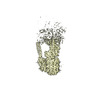
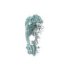
| Structure viewer | Molecule: MolmilJmol/JSmol |
|---|
- Downloads & links
-Download
| PDBx/mmCIF format | 8ki3.cif.gz | 845.2 KB | Display | PDBx/mmCIF format |
|---|---|---|---|---|
| PDB format | pdb8ki3.ent.gz | 696.5 KB | Display | PDB format |
| PDBx/mmJSON format | 8ki3.json.gz | Tree view | PDBx/mmJSON format | |
| Others |  Other downloads Other downloads |
-Validation report
| Arichive directory | https://data.pdbj.org/pub/pdb/validation_reports/ki/8ki3ftp://data.pdbj.org/pub/pdb/validation_reports/ki/8ki3 | HTTPS FTP |
|---|
-Related structure data
| Related structure data |  37251MC 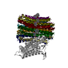 8j57C 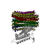 8j58C 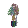 8jr0C 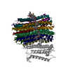 8jr1C 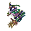 8khfC C: citing same article ( M: map data used to model this data |
|---|---|
| Similar structure data |


-Links
 PDBj
PDBj


- Assembly
| Deposited unit | 
|
|---|---|
| 1 |
|
-Components
-ATP synthase subunit ... , 12 types, 16 molecules ABCDEFGOHIMNPRST
| #1: Protein | / ATP synthase F1 subunit alpha Mass: 55276.160 Da / Num. of mol.: 3 / Source method: isolated from a natural source / Source: (natural) Homo sapiens (human) / References: UniProt: P25705#2: Protein | Mass: 51821.965 Da / Num. of mol.: 3 / Source method: isolated from a natural source / Source: (natural) Homo sapiens (human) / References: UniProt: P06576#3: Protein | | / ATP synthase F1 subunit gamma / F-ATPase gamma subunitMass: 30207.752 Da / Num. of mol.: 1 / Source method: isolated from a natural source / Source: (natural) Homo sapiens (human) / References: UniProt: P36542#5: Protein | | / ATP synthase peripheral stalk subunit OSCP / Oligomycin sensitivity conferral protein / OSCPMass: 20904.488 Da / Num. of mol.: 1 / Source method: isolated from a natural source / Source: (natural) Homo sapiens (human) / References: UniProt: P48047#7: Protein | | / ATP synthase F1 subunit delta / F-ATPase delta subunitMass: 15029.817 Da / Num. of mol.: 1 / Source method: isolated from a natural source / Source: (natural) Homo sapiens (human) / References: UniProt: P30049#8: Protein | | / ATPase subunit epsilon / ATP synthase F1 subunit epsilonMass: 5790.779 Da / Num. of mol.: 1 / Source method: isolated from a natural source / Source: (natural) Homo sapiens (human) / References: UniProt: P56381#10: Protein | | / ATPase subunit d / ATP synthase peripheral stalk subunit dMass: 18383.982 Da / Num. of mol.: 1 / Source method: isolated from a natural source / Source: (natural) Homo sapiens (human) / References: UniProt: O75947#11: Protein | | / F-ATPase protein 6Mass: 24833.102 Da / Num. of mol.: 1 / Source method: isolated from a natural source / Source: (natural) Homo sapiens (human) / References: UniProt: P00846#12: Protein | | / 6.8 kDa mitochondrial proteolipid protein / MLQ / ATP synthase membrane subunit 6.8PLMass: 6673.053 Da / Num. of mol.: 1 / Source method: isolated from a natural source / Source: (natural) Homo sapiens (human) / References: UniProt: P56378#14: Protein | | / ATP synthase membrane subunit fMass: 10804.686 Da / Num. of mol.: 1 / Source method: isolated from a natural source / Source: (natural) Homo sapiens (human) / References: UniProt: P56134#15: Protein | | / ATPase subunit g / ATP synthase membrane subunit gMass: 11309.226 Da / Num. of mol.: 1 / Source method: isolated from a natural source / Source: (natural) Homo sapiens (human) / References: UniProt: O75964#16: Protein | | / ATPase subunit e / ATP synthase membrane subunit eMass: 7947.215 Da / Num. of mol.: 1 / Source method: isolated from a natural source / Source: (natural) Homo sapiens (human) / References: UniProt: P56385 |
|---|
-Protein , 3 types, 3 molecules JQL
| #4: Protein | Mass: 9540.627 Da / Num. of mol.: 1 / Source method: isolated from a natural source / Source: (natural) Homo sapiens (human) / References: UniProt: Q9UII2 |
|---|---|
| #13: Protein | / A6L / F-ATPase subunit 8 Mass: 8000.634 Da / Num. of mol.: 1 / Source method: isolated from a natural source / Source: (natural) Homo sapiens (human) / References: UniProt: P03928 |
| #17: Protein | Mass: 12606.499 Da / Num. of mol.: 1 / Source method: isolated from a natural source / Source: (natural) Homo sapiens (human) / References: UniProt: P18859 |
-ATP synthase F(0) complex subunit ... , 2 types, 9 molecules 12345678K
| #6: Protein | Mass: 7610.954 Da / Num. of mol.: 8 / Source method: isolated from a natural source / Source: (natural) Homo sapiens (human) / References: UniProt: P05496#9: Protein | | Mass: 24658.586 Da / Num. of mol.: 1 / Source method: isolated from a natural source / Source: (natural) Homo sapiens (human) / References: UniProt: P24539 |
|---|
-Non-polymers , 4 types, 11 molecules 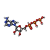


| #18: Chemical | Adenosine triphosphate Mass: 507.181 Da / Num. of mol.: 3 / Source method: obtained synthetically / Formula: C10H16N5O13P3 / Feature type: SUBJECT OF INVESTIGATION / Comment: ATP, energy-carrying molecule*YM Mass: 507.181 Da / Num. of mol.: 3 / Source method: obtained synthetically / Formula: C10H16N5O13P3 / Feature type: SUBJECT OF INVESTIGATION / Comment: ATP, energy-carrying molecule*YM#19: Chemical | ChemComp-MG /  Mass: 24.305 Da / Num. of mol.: 5 / Source method: obtained synthetically / Formula: Mg / Feature type: SUBJECT OF INVESTIGATION Mass: 24.305 Da / Num. of mol.: 5 / Source method: obtained synthetically / Formula: Mg / Feature type: SUBJECT OF INVESTIGATION#20: Chemical | Adenosine diphosphate Mass: 427.201 Da / Num. of mol.: 2 / Source method: obtained synthetically / Formula: C10H15N5O10P2 / Feature type: SUBJECT OF INVESTIGATION / Comment: ADP, energy-carrying molecule*YM Mass: 427.201 Da / Num. of mol.: 2 / Source method: obtained synthetically / Formula: C10H15N5O10P2 / Feature type: SUBJECT OF INVESTIGATION / Comment: ADP, energy-carrying molecule*YM#21: Chemical | ChemComp-BQ1 / | Bedaquiline Mass: 555.505 Da / Num. of mol.: 1 / Source method: obtained synthetically / Formula: C32H31BrN2O2 / Feature type: SUBJECT OF INVESTIGATION / Comment: medication, antibiotic*YM Mass: 555.505 Da / Num. of mol.: 1 / Source method: obtained synthetically / Formula: C32H31BrN2O2 / Feature type: SUBJECT OF INVESTIGATION / Comment: medication, antibiotic*YM |
|---|
-Details
| Has ligand of interest | Y |
|---|
-Experimental details
-Experiment
| Experiment | Method: ELECTRON MICROSCOPY |
|---|---|
| EM experiment | Aggregation state: PARTICLE / 3D reconstruction method: single particle reconstruction |
- Sample preparation
| Component | Name: human ATP synthase / Type: COMPLEX / Entity ID: #1-#17 / Source: NATURAL |
|---|---|
| Source (natural) | Organism: Homo sapiens (human) |
| Source (recombinant) | Organism: Homo sapiens (human) |
| Buffer solution | pH: 7.4 |
| Specimen | Embedding applied: NO / Shadowing applied: NO / Staining applied: NO / Vitrification applied: YES |
| Vitrification | Cryogen name: ETHANE |
- Electron microscopy imaging
| Experimental equipment |  Model: Titan Krios / Image courtesy: FEI Company |
|---|---|
| Microscopy | Model: FEI TITAN KRIOS |
| Electron gun | Electron source: FIELD EMISSION GUN / Accelerating voltage: 300 kV / Illumination mode: FLOOD BEAM |
| Electron lens | Mode: BRIGHT FIELDBright-field microscopy / Nominal defocus max: 2400 nm / Nominal defocus min: 1200 nm |
| Image recording | Electron dose: 50 e/Å2 / Film or detector model: FEI FALCON IV (4k x 4k) |
- Processing
| CTF correction | Type: PHASE FLIPPING AND AMPLITUDE CORRECTION | ||||||||||||||||||||||||
|---|---|---|---|---|---|---|---|---|---|---|---|---|---|---|---|---|---|---|---|---|---|---|---|---|---|
| 3D reconstruction | Resolution: 2.89 Å / Resolution method: FSC 0.143 CUT-OFF / Num. of particles: 84037 / Symmetry type: POINT | ||||||||||||||||||||||||
| Refine LS restraints |
|
