 Movie
Movie Controller


+ Open data

- Basic information
| Entry | Database: PDB / ID: 8imz | |||||||||
|---|---|---|---|---|---|---|---|---|---|---|
| Title | Cryo-EM structure of mouse Piezo1-MDFIC complex (composite map) | |||||||||
 Components Components |
| |||||||||
 Keywords Keywords |  MEMBRANE PROTEIN / Piezo1 complex / mechanosensation / mechanotransduction MEMBRANE PROTEIN / Piezo1 complex / mechanosensation / mechanotransduction | |||||||||
| Function / homology |  Function and homology information Function and homology informationmechanosensitive monoatomic cation channel activity / positive regulation of viral transcription / positive regulation of cell-cell adhesion mediated by integrin / detection of mechanical stimulus / mechanosensitive monoatomic ion channel activity / positive regulation of integrin activation / Tat protein binding / regulation of Wnt signaling pathway / negative regulation of protein import into nucleus / positive regulation of myotube differentiation ...mechanosensitive monoatomic cation channel activity / positive regulation of viral transcription / positive regulation of cell-cell adhesion mediated by integrin / detection of mechanical stimulus / mechanosensitive monoatomic ion channel activity / positive regulation of integrin activation / Tat protein binding / regulation of Wnt signaling pathway / negative regulation of protein import into nucleus / positive regulation of myotube differentiation / lamellipodium membrane / monoatomic cation transport / regulation of JNK cascade / monoatomic cation channel activity / regulation of membrane potential / endoplasmic reticulum-Golgi intermediate compartment membrane / cyclin binding / cellular response to mechanical stimulus / DNA-binding transcription factor binding / negative regulation of DNA-templated transcription / endoplasmic reticulum membrane / nucleolus / positive regulation of DNA-templated transcription / endoplasmic reticulum / extracellular region / identical protein binding / nucleus / plasma membrane / cytoplasmSimilarity search - Function | |||||||||
| Biological species |  Mus musculus (house mouse) Mus musculus (house mouse) | |||||||||
| Method | ELECTRON MICROSCOPY / single particle reconstruction / cryo EM / Resolution: 3.66 Å | |||||||||
 Authors Authors | Zhou, Z. / Ma, X. / Lin, Y. / Cheng, D. / Bavi, N. / Li, J.V. / Sutton, D. / Yao, M. / Harvey, N. / Corry, B. ...Zhou, Z. / Ma, X. / Lin, Y. / Cheng, D. / Bavi, N. / Li, J.V. / Sutton, D. / Yao, M. / Harvey, N. / Corry, B. / Zhang, Y. / Cox, C.D. | |||||||||
| Funding support |  Australia, Australia,  China, 2items China, 2items
| |||||||||
 Citation Citation | Journal: Science / Year: 2023 Title: MyoD-family inhibitor proteins act as auxiliary subunits of Piezo channels. Authors: Zijing Zhou / Xiaonuo Ma / Yiechang Lin / Delfine Cheng / Navid Bavi / Genevieve A Secker / Jinyuan Vero Li / Vaibhao Janbandhu / Drew L Sutton / Hamish S Scott / Mingxi Yao / Richard P ...Authors: Zijing Zhou / Xiaonuo Ma / Yiechang Lin / Delfine Cheng / Navid Bavi / Genevieve A Secker / Jinyuan Vero Li / Vaibhao Janbandhu / Drew L Sutton / Hamish S Scott / Mingxi Yao / Richard P Harvey / Natasha L Harvey / Ben Corry / Yixiao Zhang / Charles D Cox /  Abstract: Piezo channels are critical cellular sensors of mechanical forces. Despite their large size, ubiquitous expression, and irreplaceable roles in an ever-growing list of physiological processes, few ...Piezo channels are critical cellular sensors of mechanical forces. Despite their large size, ubiquitous expression, and irreplaceable roles in an ever-growing list of physiological processes, few Piezo channel-binding proteins have emerged. In this work, we found that MyoD (myoblast determination)-family inhibitor proteins (MDFIC and MDFI) are PIEZO1/2 interacting partners. These transcriptional regulators bind to PIEZO1/2 channels, regulating channel inactivation. Using single-particle cryogenic electron microscopy, we mapped the interaction site in MDFIC to a lipidated, C-terminal helix that inserts laterally into the PIEZO1 pore module. These Piezo-interacting proteins fit all the criteria for auxiliary subunits, contribute to explaining the vastly different gating kinetics of endogenous Piezo channels observed in many cell types, and elucidate mechanisms potentially involved in human lymphatic vascular disease. | |||||||||
| History |
|
- Structure visualization
| Structure viewer | Molecule: MolmilJmol/JSmol |
|---|
- Downloads & links
-Download
| PDBx/mmCIF format | 8imz.cif.gz | 724.3 KB | Display | PDBx/mmCIF format |
|---|---|---|---|---|
| PDB format | pdb8imz.ent.gz | 547.5 KB | Display | PDB format |
| PDBx/mmJSON format | 8imz.json.gz | Tree view | PDBx/mmJSON format | |
| Others |  Other downloads Other downloads |
-Validation report
| Arichive directory | https://data.pdbj.org/pub/pdb/validation_reports/im/8imzftp://data.pdbj.org/pub/pdb/validation_reports/im/8imz | HTTPS FTP |
|---|
-Related structure data
| Related structure data | 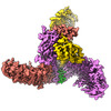 35577MC M: map data used to model this data C: citing same article ( |
|---|---|
| Similar structure data |
-Links
 PDBj
PDBj
- Assembly
| Deposited unit | 
|
|---|---|
| 1 |
|
-Components
| #1: Protein | Mass: 292320.656 Da / Num. of mol.: 3 Source method: isolated from a genetically manipulated source Source: (gene. exp.) Mus musculus (house mouse) / Gene: Piezo1, Fam38a / Production host:  Homo sapiens (human) / References: UniProt: E2JF22 Homo sapiens (human) / References: UniProt: E2JF22#2: Protein | Mass: 26122.732 Da / Num. of mol.: 3 Source method: isolated from a genetically manipulated source Source: (gene. exp.) Mus musculus (house mouse) / Gene: Mdfic, Kdt1 / Production host: Homo sapiens (human) / References: UniProt: Q8BX65 |
|---|
-Experimental details
-Experiment
| Experiment | Method: ELECTRON MICROSCOPY |
|---|---|
| EM experiment | Aggregation state: PARTICLE / 3D reconstruction method: single particle reconstruction |
- Sample preparation
| Component | Name: Piezo1 in complex with MDFIC / Type: COMPLEX / Entity ID: all / Source: MULTIPLE SOURCES |
|---|---|
| Source (natural) | Organism: Mus musculus (house mouse) |
| Source (recombinant) | Organism: Homo sapiens (human) |
| Buffer solution | pH: 7.9 |
| Specimen | Embedding applied: NO / Shadowing applied: NO / Staining applied: NO / Vitrification applied: YES |
| Vitrification | Cryogen name: ETHANE |
- Electron microscopy imaging
| Experimental equipment |  Model: Titan Krios / Image courtesy: FEI Company |
|---|---|
| Microscopy | Model: FEI TITAN KRIOS |
| Electron gun | Electron source: FIELD EMISSION GUN / Accelerating voltage: 300 kV / Illumination mode: FLOOD BEAM |
| Electron lens | Mode: BRIGHT FIELDBright-field microscopy / Nominal defocus max: 2400 nm / Nominal defocus min: 1200 nm |
| Image recording | Electron dose: 49.4 e/Å2 / Film or detector model: GATAN K3 BIOQUANTUM (6k x 4k) |
- Processing
| Software | Name: PHENIX / Version: 1.18.2_3874: / Classification: refinement | ||||||||||||||||||||||||
|---|---|---|---|---|---|---|---|---|---|---|---|---|---|---|---|---|---|---|---|---|---|---|---|---|---|
| CTF correction | Type: PHASE FLIPPING AND AMPLITUDE CORRECTION | ||||||||||||||||||||||||
| 3D reconstruction | Resolution: 3.66 Å / Resolution method: FSC 0.143 CUT-OFF / Num. of particles: 102644 / Symmetry type: POINT | ||||||||||||||||||||||||
| Refine LS restraints |
|
