 Movie
Movie Controller


+ Open data

- Basic information
| Entry | Database: PDB / ID: 8i2z | ||||||
|---|---|---|---|---|---|---|---|
| Title | Cryo-EM structure of the zeaxanthin-bound kin4B8 | ||||||
 Components Components | Xanthorhodopsin | ||||||
 Keywords Keywords |  PROTON TRANSPORT / Rhodopsin / Cryo-EM / anntena PROTON TRANSPORT / Rhodopsin / Cryo-EM / anntena | ||||||
| Function / homology | Zeaxanthin / RETINAL / Xanthorhodopsin Function and homology information Function and homology information | ||||||
| Biological species |  uncultured Bdellovibrionales bacterium (environmental samples) uncultured Bdellovibrionales bacterium (environmental samples) | ||||||
| Method | ELECTRON MICROSCOPY / single particle reconstruction / cryo EM / Resolution: 2.3 Å | ||||||
 Authors Authors | Murakoshi, S. / Chazan, A. / Shihoya, W. / Beja, O. / Nureki, O. | ||||||
| Funding support |  Japan, 1items Japan, 1items
| ||||||
 Citation Citation | Journal: Nature / Year: 2023 Title: Phototrophy by antenna-containing rhodopsin pumps in aquatic environments. Authors: Ariel Chazan / Ishita Das / Takayoshi Fujiwara / Shunya Murakoshi / Andrey Rozenberg / Ana Molina-Márquez / Fumiya K Sano / Tatsuki Tanaka / Patricia Gómez-Villegas / Shirley Larom / Alina ...Authors: Ariel Chazan / Ishita Das / Takayoshi Fujiwara / Shunya Murakoshi / Andrey Rozenberg / Ana Molina-Márquez / Fumiya K Sano / Tatsuki Tanaka / Patricia Gómez-Villegas / Shirley Larom / Alina Pushkarev / Partha Malakar / Masumi Hasegawa / Yuya Tsukamoto / Tomohiro Ishizuka / Masae Konno / Takashi Nagata / Yosuke Mizuno / Kota Katayama / Rei Abe-Yoshizumi / Sanford Ruhman / Keiichi Inoue / Hideki Kandori / Rosa León / Wataru Shihoya / Susumu Yoshizawa / Mordechai Sheves / Osamu Nureki / Oded Béjà /    Abstract: Energy transfer from light-harvesting ketocarotenoids to the light-driven proton pump xanthorhodopsins has been previously demonstrated in two unique cases: an extreme halophilic bacterium and a ...Energy transfer from light-harvesting ketocarotenoids to the light-driven proton pump xanthorhodopsins has been previously demonstrated in two unique cases: an extreme halophilic bacterium and a terrestrial cyanobacterium. Attempts to find carotenoids that bind and transfer energy to abundant rhodopsin proton pumps from marine photoheterotrophs have thus far failed. Here we detected light energy transfer from the widespread hydroxylated carotenoids zeaxanthin and lutein to the retinal moiety of xanthorhodopsins and proteorhodopsins using functional metagenomics combined with chromophore extraction from the environment. The light-harvesting carotenoids transfer up to 42% of the harvested energy in the violet- or blue-light range to the green-light absorbing retinal chromophore. Our data suggest that these antennas may have a substantial effect on rhodopsin phototrophy in the world's lakes, seas and oceans. However, the functional implications of our findings are yet to be discovered. | ||||||
| History |
|
- Structure visualization
| Structure viewer | Molecule: MolmilJmol/JSmol |
|---|
- Downloads & links
-Download
| PDBx/mmCIF format | 8i2z.cif.gz | 61.3 KB | Display | PDBx/mmCIF format |
|---|---|---|---|---|
| PDB format | pdb8i2z.ent.gz | 44.9 KB | Display | PDB format |
| PDBx/mmJSON format | 8i2z.json.gz | Tree view | PDBx/mmJSON format | |
| Others |  Other downloads Other downloads |
-Validation report
| Arichive directory | https://data.pdbj.org/pub/pdb/validation_reports/i2/8i2zftp://data.pdbj.org/pub/pdb/validation_reports/i2/8i2z | HTTPS FTP |
|---|
-Related structure data
| Related structure data | 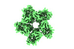 35143MC  7ytbC M: map data used to model this data C: citing same article ( |
|---|---|
| Similar structure data |
-Links
 PDBj
PDBj


- Assembly
| Deposited unit | 
|
|---|---|
| 1 | x 5
|
| 2 |
|
| 3 | 
|
| Symmetry | Point symmetry: (Schoenflies symbol: C5 (5 fold cyclic)) |
-Components
| #1: Protein | Mass: 28745.842 Da / Num. of mol.: 1 Source method: isolated from a genetically manipulated source Source: (gene. exp.) uncultured Bdellovibrionales bacterium (environmental samples)Gene: tmp_000014 / Production host: Escherichia coli (E. coli) / References: UniProt: A0A977XLG0 |
|---|---|
| #2: Chemical | ChemComp-RET / Retinal  Mass: 284.436 Da / Num. of mol.: 1 / Source method: obtained synthetically / Formula: C20H28O / Feature type: SUBJECT OF INVESTIGATION Mass: 284.436 Da / Num. of mol.: 1 / Source method: obtained synthetically / Formula: C20H28O / Feature type: SUBJECT OF INVESTIGATION |
| #3: Chemical | ChemComp-K3I /   Mass: 568.871 Da / Num. of mol.: 1 / Source method: obtained synthetically / Formula: C40H56O2 / Feature type: SUBJECT OF INVESTIGATION Mass: 568.871 Da / Num. of mol.: 1 / Source method: obtained synthetically / Formula: C40H56O2 / Feature type: SUBJECT OF INVESTIGATION |
| #4: Water | ChemComp-HOH / Water Mass: 18.015 Da / Num. of mol.: 33 / Source method: isolated from a natural source / Formula: H2O Mass: 18.015 Da / Num. of mol.: 33 / Source method: isolated from a natural source / Formula: H2O |
| Has ligand of interest | Y |
-Experimental details
-Experiment
| Experiment | Method: ELECTRON MICROSCOPY |
|---|---|
| EM experiment | Aggregation state: PARTICLE / 3D reconstruction method: single particle reconstruction |
- Sample preparation
| Component | Name: Kin4B8 / Type: ORGANELLE OR CELLULAR COMPONENT / Entity ID: #1 / Source: RECOMBINANT | |||||||||||||||
|---|---|---|---|---|---|---|---|---|---|---|---|---|---|---|---|---|
| Molecular weight | Experimental value: NO | |||||||||||||||
| Source (natural) | Organism: uncultured Bdellovibrionales bacterium (environmental samples) | |||||||||||||||
| Source (recombinant) | Organism: Escherichia coli (E. coli) | |||||||||||||||
| Buffer solution | pH: 8 / Details: 150 mM NaCl, 20 mM Tris-HCl | |||||||||||||||
| Buffer component |
| |||||||||||||||
| Specimen | Embedding applied: NO / Shadowing applied: NO / Staining applied: NO / Vitrification applied: YES | |||||||||||||||
| Specimen support | Grid material: GOLD / Grid mesh size: 300 divisions/in. / Grid type: Quantifoil R1.2/1.3 | |||||||||||||||
| Vitrification | Cryogen name: ETHANE / Humidity: 100 % / Chamber temperature: 277 K / Details: Vitrification carried out in ethane atmosphere. |
- Electron microscopy imaging
| Experimental equipment |  Model: Titan Krios / Image courtesy: FEI Company |
|---|---|
| Microscopy | Model: TFS KRIOS |
| Electron gun | Electron source: FIELD EMISSION GUN / Accelerating voltage: 300 kV / Illumination mode: FLOOD BEAM |
| Electron lens | Mode: BRIGHT FIELDBright-field microscopy / Nominal defocus max: 1600 nm / Nominal defocus min: 600 nm |
| Image recording | Electron dose: 50.14 e/Å2 / Film or detector model: GATAN K3 (6k x 4k) |
- Processing
| Software | Name: REFMAC / Version: 5.8.0267 / Classification: refinement | ||||||||||||||||||||||||||||||||||||||||||||||||||||||||||||||||||||||||||||||||||||||||||||||||||||||||||
|---|---|---|---|---|---|---|---|---|---|---|---|---|---|---|---|---|---|---|---|---|---|---|---|---|---|---|---|---|---|---|---|---|---|---|---|---|---|---|---|---|---|---|---|---|---|---|---|---|---|---|---|---|---|---|---|---|---|---|---|---|---|---|---|---|---|---|---|---|---|---|---|---|---|---|---|---|---|---|---|---|---|---|---|---|---|---|---|---|---|---|---|---|---|---|---|---|---|---|---|---|---|---|---|---|---|---|---|
| EM software |
| ||||||||||||||||||||||||||||||||||||||||||||||||||||||||||||||||||||||||||||||||||||||||||||||||||||||||||
| CTF correction | Type: PHASE FLIPPING AND AMPLITUDE CORRECTION | ||||||||||||||||||||||||||||||||||||||||||||||||||||||||||||||||||||||||||||||||||||||||||||||||||||||||||
| 3D reconstruction | Resolution: 2.3 Å / Resolution method: FSC 0.143 CUT-OFF / Num. of particles: 4337540 / Symmetry type: POINT | ||||||||||||||||||||||||||||||||||||||||||||||||||||||||||||||||||||||||||||||||||||||||||||||||||||||||||
| Refinement | Resolution: 2→107.9 Å / Cor.coef. Fo:Fc: 0.899 / SU B: 3.934 / SU ML: 0.087 / ESU R: 0.05 Stereochemistry target values: MAXIMUM LIKELIHOOD WITH PHASES Details: HYDROGENS HAVE BEEN ADDED IN THE RIDING POSITIONS
| ||||||||||||||||||||||||||||||||||||||||||||||||||||||||||||||||||||||||||||||||||||||||||||||||||||||||||
| Solvent computation | Solvent model: PARAMETERS FOR MASK CACLULATION | ||||||||||||||||||||||||||||||||||||||||||||||||||||||||||||||||||||||||||||||||||||||||||||||||||||||||||
| Displacement parameters | Biso mean: 86.425 Å2 | ||||||||||||||||||||||||||||||||||||||||||||||||||||||||||||||||||||||||||||||||||||||||||||||||||||||||||
| Refinement step | Cycle: 1 / Total: 2050 | ||||||||||||||||||||||||||||||||||||||||||||||||||||||||||||||||||||||||||||||||||||||||||||||||||||||||||
| Refine LS restraints |
|