 Movie
Movie Controller

[English] 日本語
 Yorodumi
- PDB-8gb3: Structure of the Mycobacterium tuberculosis Hsp70 protein DnaK bo... -
+ Open data

- Basic information
| Entry | Database: PDB / ID: 8gb3 | ||||||
|---|---|---|---|---|---|---|---|
| Title | Structure of the Mycobacterium tuberculosis Hsp70 protein DnaK bound to the nucleotide exchange factor GrpE | ||||||
 Components Components |
| ||||||
 Keywords Keywords |  CHAPERONE / heat shock protein 70 / nucleotide exchange factor / protein folding and refolding CHAPERONE / heat shock protein 70 / nucleotide exchange factor / protein folding and refolding | ||||||
| Function / homology |  Function and homology information Function and homology informationadenyl-nucleotide exchange factor activity / ATP-dependent protein folding chaperone / unfolded protein binding / protein folding / protein-folding chaperone binding / hydrolase activity / protein homodimerization activity / ATP binding / cytoplasmSimilarity search - Function | ||||||
| Biological species |  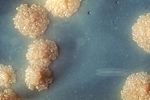 Mycobacterium tuberculosis (bacteria) Mycobacterium tuberculosis (bacteria) | ||||||
| Method | ELECTRON MICROSCOPY / single particle reconstruction / cryo EM / Resolution: 3.7 Å | ||||||
 Authors Authors | Xiao, X. / Li, H. | ||||||
| Funding support |  United States, 1items United States, 1items
| ||||||
 Citation Citation | Journal: Nat Commun / Year: 2024 Title: Structure of the M. tuberculosis DnaK-GrpE complex reveals how key DnaK roles are controlled. Authors: Xiansha Xiao / Allison Fay / Pablo Santos Molina / Amanda Kovach / Michael S Glickman / Huilin Li / Abstract: The molecular chaperone DnaK is essential for viability of Mycobacterium tuberculosis (Mtb). DnaK hydrolyzes ATP to fold substrates, and the resulting ADP is exchanged for ATP by the nucleotide ...The molecular chaperone DnaK is essential for viability of Mycobacterium tuberculosis (Mtb). DnaK hydrolyzes ATP to fold substrates, and the resulting ADP is exchanged for ATP by the nucleotide exchange factor GrpE. It has been unclear how GrpE couples DnaK's nucleotide exchange with substrate release. Here we report a cryo-EM analysis of GrpE bound to an intact Mtb DnaK, revealing an asymmetric 1:2 DnaK-GrpE complex. The GrpE dimer ratchets to modulate both DnaK nucleotide-binding domain and the substrate-binding domain. We further show that the disordered GrpE N-terminus is critical for substrate release, and that the DnaK-GrpE interface is essential for protein folding activity both in vitro and in vivo. Therefore, the Mtb GrpE dimer allosterically regulates DnaK to concomitantly release ADP in the nucleotide-binding domain and substrate peptide in the substrate-binding domain. | ||||||
| History |
|
- Structure visualization
| Structure viewer | Molecule: MolmilJmol/JSmol |
|---|
- Downloads & links
-Download
| PDBx/mmCIF format | 8gb3.cif.gz | 141.4 KB | Display | PDBx/mmCIF format |
|---|---|---|---|---|
| PDB format | pdb8gb3.ent.gz | 106.4 KB | Display | PDB format |
| PDBx/mmJSON format | 8gb3.json.gz | Tree view | PDBx/mmJSON format | |
| Others |  Other downloads Other downloads |
-Validation report
| Arichive directory | https://data.pdbj.org/pub/pdb/validation_reports/gb/8gb3ftp://data.pdbj.org/pub/pdb/validation_reports/gb/8gb3 | HTTPS FTP |
|---|
-Related structure data
| Related structure data |  29912MC M: map data used to model this data C: citing same article ( |
|---|---|
| Similar structure data |


-Links
 PDBj
PDBj
- Assembly
| Deposited unit | 
|
|---|---|
| 1 |
|
-Components
| #1: Protein | / HSP70 / Heat shock 70 kDa protein / Heat shock protein 70 Mass: 66910.680 Da / Num. of mol.: 1 Source method: isolated from a genetically manipulated source Source: (gene. exp.) Mycobacterium tuberculosis (bacteria)Gene: dnaK_2, dnaK, E5M05_19850, ERS007703_00955, ERS023446_02581, ERS027651_01905, FCN16_12395, SAMEA2682864_01182, SAMEA2683035_02658 Production host: Escherichia coli (E. coli) / References: UniProt: A0A045JRR0 |
|---|---|
| #2: Protein | Mass: 24559.555 Da / Num. of mol.: 2 Source method: isolated from a genetically manipulated source Source: (gene. exp.) Mycobacterium tuberculosis (bacteria)Gene: grpE, E5M05_19845, E5M52_19255, E5M78_19290, ERS007681_03281, ERS007703_00954, ERS024276_02384, SAMEA2683035_02659 Production host: Escherichia coli (E. coli) / References: UniProt: A0A045J399 |
-Experimental details
-Experiment
| Experiment | Method: ELECTRON MICROSCOPY |
|---|---|
| EM experiment | Aggregation state: PARTICLE / 3D reconstruction method: single particle reconstruction |
- Sample preparation
| Component | Name: Binary complex of DnaK with nucleotide exchange factor GrpE Type: COMPLEX / Entity ID: all / Source: RECOMBINANT |
|---|---|
| Source (natural) | Organism: Mycobacterium tuberculosis (bacteria) |
| Source (recombinant) | Organism: Escherichia coli (E. coli) |
| Buffer solution | pH: 8 |
| Specimen | Embedding applied: NO / Shadowing applied: NO / Staining applied: NO / Vitrification applied: YES |
| Specimen support | Grid material: COPPER / Grid mesh size: 300 divisions/in. / Grid type: Quantifoil R2/1 |
| Vitrification | Instrument: FEI VITROBOT MARK II / Cryogen name: ETHANE / Humidity: 100 % / Chamber temperature: 283.15 K |
- Electron microscopy imaging
| Experimental equipment |  Model: Titan Krios / Image courtesy: FEI Company |
|---|---|
| Microscopy | Model: FEI TITAN KRIOS |
| Electron gun | Electron source: FIELD EMISSION GUN / Accelerating voltage: 300 kV / Illumination mode: FLOOD BEAM |
| Electron lens | Mode: BRIGHT FIELDBright-field microscopy / Nominal magnification: 105000 X / Nominal defocus max: 1800 nm / Nominal defocus min: 1300 nm / Cs: 2.7 mm / C2 aperture diameter: 100 µm / Alignment procedure: COMA FREE |
| Specimen holder | Cryogen: NITROGEN / Specimen holder model: FEI TITAN KRIOS AUTOGRID HOLDER |
| Image recording | Average exposure time: 1.5 sec. / Electron dose: 66 e/Å2 / Detector mode: SUPER-RESOLUTION / Film or detector model: GATAN K3 BIOQUANTUM (6k x 4k) / Num. of real images: 15720 |
| EM imaging optics | Energyfilter name: GIF Bioquantum / Energyfilter slit width: 20 eV |
| Image scans | Movie frames/image: 30 |
- Processing
| Software | Name: PHENIX / Version: 1.20.1_4487: / Classification: refinement | ||||||||||||||||||||||||||||||||||||||||
|---|---|---|---|---|---|---|---|---|---|---|---|---|---|---|---|---|---|---|---|---|---|---|---|---|---|---|---|---|---|---|---|---|---|---|---|---|---|---|---|---|---|
| EM software |
| ||||||||||||||||||||||||||||||||||||||||
| CTF correction | Type: PHASE FLIPPING AND AMPLITUDE CORRECTION | ||||||||||||||||||||||||||||||||||||||||
| Particle selection | Num. of particles selected: 9973114 | ||||||||||||||||||||||||||||||||||||||||
| 3D reconstruction | Resolution: 3.7 Å / Resolution method: FSC 0.143 CUT-OFF / Num. of particles: 240737 / Symmetry type: POINT | ||||||||||||||||||||||||||||||||||||||||
| Refine LS restraints |
|
