 Movie
Movie Controller


+ Open data

- Basic information
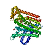
| Entry | Database: PDB / ID: 8f8l | ||||||
|---|---|---|---|---|---|---|---|
| Title | The structure of Rv2173 from M. tuberculosis with DMAP bound | ||||||
 Components Components | (2E,6E)-farnesyl diphosphate synthase | ||||||
 Keywords Keywords |  TRANSFERASE / Rv2173 / M. tuberculosis / Isoprenyl diphosphate synthase / dimethylallyl diphosphate (DMAP) bound TRANSFERASE / Rv2173 / M. tuberculosis / Isoprenyl diphosphate synthase / dimethylallyl diphosphate (DMAP) bound | ||||||
| Function / homology |  Function and homology information Function and homology informationgeranyl diphosphate biosynthetic process / dimethylallyltranstransferase / (2E,6E)-farnesyl diphosphate synthase / geranyltranstransferase activity / prenyltransferase activity / dimethylallyltranstransferase activity / isoprenoid biosynthetic process / metal ion bindingSimilarity search - Function | ||||||
| Biological species |   Mycobacterium tuberculosis (bacteria) Mycobacterium tuberculosis (bacteria) | ||||||
| Method | X-RAY DIFFRACTION / SYNCHROTRON / MOLECULAR REPLACEMENT / Resolution: 2.2 Å | ||||||
 Authors Authors | Johnston, J.M. / Allison, T.M. / Titterington, J. | ||||||
| Funding support |  New Zealand, 1items New Zealand, 1items
| ||||||
 Citation Citation | Journal: To be Published Title: The structure of Rv2173 from M. tuberculosis in APO-, IPP-, and DMAP-bound forms. Authors: Johnston, J.M. / Allison, T.M. / Titterington, J. / Beasley, C.P.H. | ||||||
| History |
|
- Structure visualization
| Structure viewer | Molecule: MolmilJmol/JSmol |
|---|
- Downloads & links
-Download
| PDBx/mmCIF format | 8f8l.cif.gz | 85.7 KB | Display | PDBx/mmCIF format |
|---|---|---|---|---|
| PDB format | pdb8f8l.ent.gz | 60.1 KB | Display | PDB format |
| PDBx/mmJSON format | 8f8l.json.gz | Tree view | PDBx/mmJSON format | |
| Others |  Other downloads Other downloads |
-Validation report
| Arichive directory | https://data.pdbj.org/pub/pdb/validation_reports/f8/8f8lftp://data.pdbj.org/pub/pdb/validation_reports/f8/8f8l | HTTPS FTP |
|---|
-Related structure data
-Links
 PDBj
PDBj
- Assembly
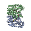
| Deposited unit | 
| ||||||||
|---|---|---|---|---|---|---|---|---|---|
| 1 |
| ||||||||
| Unit cell |
|
-Components
| #1: Protein | ( Mass: 41981.621 Da / Num. of mol.: 1 Source method: isolated from a genetically manipulated source Source: (gene. exp.) Mycobacterium tuberculosis (bacteria) / Gene: idsA2, Rv2173 / Production host: Escherichia coli BL21(DE3) (bacteria)References: UniProt: O53507, (2E,6E)-farnesyl diphosphate synthase, dimethylallyltranstransferase | ||||
|---|---|---|---|---|---|
| #2: Chemical | ChemComp-DMA / Dimethylallyl pyrophosphate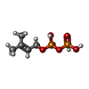  Mass: 246.092 Da / Num. of mol.: 1 / Source method: obtained synthetically / Formula: C5H12O7P2 / Feature type: SUBJECT OF INVESTIGATION Mass: 246.092 Da / Num. of mol.: 1 / Source method: obtained synthetically / Formula: C5H12O7P2 / Feature type: SUBJECT OF INVESTIGATION | ||||
| #3: Chemical | ChemComp-GOL / Glycerol  Mass: 92.094 Da / Num. of mol.: 1 / Source method: obtained synthetically / Formula: C3H8O3 Mass: 92.094 Da / Num. of mol.: 1 / Source method: obtained synthetically / Formula: C3H8O3 | ||||
| #4: Chemical |   Mass: 40.078 Da / Num. of mol.: 2 / Source method: obtained synthetically / Formula: Ca / Feature type: SUBJECT OF INVESTIGATION Mass: 40.078 Da / Num. of mol.: 2 / Source method: obtained synthetically / Formula: Ca / Feature type: SUBJECT OF INVESTIGATION#5: Water | ChemComp-HOH / | Water Mass: 18.015 Da / Num. of mol.: 138 / Source method: isolated from a natural source / Formula: H2O Mass: 18.015 Da / Num. of mol.: 138 / Source method: isolated from a natural source / Formula: H2OHas ligand of interest | Y | |
-Experimental details
-Experiment
| Experiment | Method: X-RAY DIFFRACTION / Number of used crystals: 1 |
|---|
- Sample preparation
| Crystal | Density Matthews: 3.14 Å3/Da / Density % sol: 60.82 % |
|---|---|
| Crystal grow | Temperature: 291 K / Method: vapor diffusion Details: 0.1 M Bicine-Tris, pH 8.3, 12.5% PEG4000, 25% glycerol, 0.03 M CA mix Ca/Mg |
-Data collection
| Diffraction | Mean temperature: 110 K / Serial crystal experiment: N |
|---|---|
| Diffraction source | Source: SYNCHROTRON / Site: Australian Synchrotron  / Beamline: MX2 / Wavelength: 0.9537 Å / Beamline: MX2 / Wavelength: 0.9537 Å |
| Detector | Type: ADSC QUANTUM 315r / Detector: CCD / Date: Jul 11, 2016 |
| Radiation | Protocol: SINGLE WAVELENGTH / Monochromatic (M) / Laue (L): M / Scattering type: x-ray |
| Radiation wavelength | Wavelength: 0.9537 Å / Relative weight: 1 |
| Reflection | Resolution: 2.2→19.58 Å / Num. obs: 26527 / % possible obs: 97.2 % / Redundancy: 13.8 % / CC1/2: 0.996 / Rmerge(I) obs: 0.229 / Rpim(I) all: 0.064 / Rrim(I) all: 0.238 / Χ2: 0.98 / Net I/σ(I): 12.6 / Num. measured all: 365883 |
| Reflection shell | Resolution: 2.2→2.27 Å / % possible obs: 82 % / Redundancy: 9.1 % / Rmerge(I) obs: 2.696 / Num. measured all: 18434 / Num. unique obs: 2016 / CC1/2: 0.238 / Rpim(I) all: 0.923 / Rrim(I) all: 2.861 / Χ2: 1.03 / Net I/σ(I) obs: 0.9 |
- Processing
| Software |
| |||||||||||||||||||||||||||||||||||||||||||||||||||||||||||||||||||||||||||||
|---|---|---|---|---|---|---|---|---|---|---|---|---|---|---|---|---|---|---|---|---|---|---|---|---|---|---|---|---|---|---|---|---|---|---|---|---|---|---|---|---|---|---|---|---|---|---|---|---|---|---|---|---|---|---|---|---|---|---|---|---|---|---|---|---|---|---|---|---|---|---|---|---|---|---|---|---|---|---|
| Refinement | Method to determine structure: MOLECULAR REPLACEMENT / Resolution: 2.2→19.58 Å / SU ML: 0.27 / Cross valid method: FREE R-VALUE / σ(F): 1.34 / Phase error: 25.52 / Stereochemistry target values: ML
| |||||||||||||||||||||||||||||||||||||||||||||||||||||||||||||||||||||||||||||
| Solvent computation | Shrinkage radii: 0.9 Å / VDW probe radii: 1.1 Å / Solvent model: FLAT BULK SOLVENT MODEL | |||||||||||||||||||||||||||||||||||||||||||||||||||||||||||||||||||||||||||||
| Refinement step | Cycle: LAST / Resolution: 2.2→19.58 Å
| |||||||||||||||||||||||||||||||||||||||||||||||||||||||||||||||||||||||||||||
| Refine LS restraints |
| |||||||||||||||||||||||||||||||||||||||||||||||||||||||||||||||||||||||||||||
| LS refinement shell |
|
