Movie
Movie Controller

[English] 日本語
 Yorodumi
- PDB-8e3i: CRYO-EM STRUCTURE OF the human MPSF IN COMPLEX WITH THE AUUAAA po... -
+ Open data

- Basic information
| Entry | Database: PDB / ID: 8e3i | ||||||
|---|---|---|---|---|---|---|---|
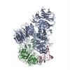
| Title | CRYO-EM STRUCTURE OF the human MPSF IN COMPLEX WITH THE AUUAAA poly(A) signal | ||||||
 Components Components |
| ||||||
 Keywords Keywords |  RNA BINDING PROTEIN / mRNA / 3'processing / polyadenylation / CPSF RNA BINDING PROTEIN / mRNA / 3'processing / polyadenylation / CPSF | ||||||
| Function / homology |  Function and homology information Function and homology informationco-transcriptional RNA 3'-end processing, cleavage and polyadenylation pathway / Inhibition of Host mRNA Processing and RNA Silencing / Processing of Intronless Pre-mRNAs / mRNA cleavage and polyadenylation specificity factor complex / mRNA 3'-UTR AU-rich region binding / collagen trimer / mRNA 3'-end processing / Transport of Mature mRNA Derived from an Intronless Transcript / postreplication repair / tRNA processing in the nucleus ...co-transcriptional RNA 3'-end processing, cleavage and polyadenylation pathway / Inhibition of Host mRNA Processing and RNA Silencing / Processing of Intronless Pre-mRNAs / mRNA cleavage and polyadenylation specificity factor complex / mRNA 3'-UTR AU-rich region binding / collagen trimer / mRNA 3'-end processing / Transport of Mature mRNA Derived from an Intronless Transcript / postreplication repair / tRNA processing in the nucleus / RNA Polymerase II Transcription Termination / : / Processing of Capped Intron-Containing Pre-mRNA / fibrillar center / mRNA processing / sequence-specific double-stranded DNA binding / spermatogenesis / intracellular membrane-bounded organelle / enzyme binding / RNA binding / zinc ion binding / nucleoplasm / nucleusSimilarity search - Function | ||||||
| Biological species |  Homo sapiens (human) Homo sapiens (human) | ||||||
| Method | ELECTRON MICROSCOPY / single particle reconstruction / cryo EM / Resolution: 2.53 Å | ||||||
 Authors Authors | Gutierrez, P.A. / Wei, J. / Sun, Y. / Tong, L. | ||||||
| Funding support |  United States, 1items United States, 1items
| ||||||
 Citation Citation | Journal: RNA / Year: 2022 Title: Molecular basis for the recognition of the AUUAAA polyadenylation signal by mPSF. Authors: Pedro A Gutierrez / Jia Wei / Yadong Sun / Liang Tong / Abstract: The polyadenylation signal (PAS) is a key sequence element for 3'-end cleavage and polyadenylation of messenger RNA precursors (pre-mRNAs). This hexanucleotide motif is recognized by the mammalian ...The polyadenylation signal (PAS) is a key sequence element for 3'-end cleavage and polyadenylation of messenger RNA precursors (pre-mRNAs). This hexanucleotide motif is recognized by the mammalian polyadenylation specificity factor (mPSF), consisting of CPSF160, WDR33, CPSF30, and Fip1 subunits. Recent studies have revealed how the AAUAAA PAS, the most frequently observed PAS, is recognized by mPSF. We report here the structure of human mPSF in complex with the AUUAAA PAS, the second most frequently identified PAS. Conformational differences are observed for the A1 and U2 nucleotides in AUUAAA compared to the A1 and A2 nucleotides in AAUAAA, while the binding modes of the remaining 4 nt are essentially identical. The 5' phosphate of U2 moves by 2.6 Å and the U2 base is placed near the six-membered ring of A2 in AAUAAA, where it makes two hydrogen bonds with zinc finger 2 (ZF2) of CPSF30, which undergoes conformational changes as well. We also attempted to determine the binding modes of two rare PAS hexamers, AAGAAA and GAUAAA, but did not observe the RNA in the cryo-electron microscopy density. The residues in CPSF30 (ZF2 and ZF3) and WDR33 that recognize PAS are disordered in these two structures. | ||||||
| History |
|
- Structure visualization
| Structure viewer | Molecule: MolmilJmol/JSmol |
|---|
- Downloads & links
-Download
| PDBx/mmCIF format | 8e3i.cif.gz | 318 KB | Display | PDBx/mmCIF format |
|---|---|---|---|---|
| PDB format | pdb8e3i.ent.gz | 245.9 KB | Display | PDB format |
| PDBx/mmJSON format | 8e3i.json.gz | Tree view | PDBx/mmJSON format | |
| Others |  Other downloads Other downloads |
-Validation report
| Arichive directory | https://data.pdbj.org/pub/pdb/validation_reports/e3/8e3iftp://data.pdbj.org/pub/pdb/validation_reports/e3/8e3i | HTTPS FTP |
|---|
-Related structure data
| Related structure data |  27866MC  8e3qC M: map data used to model this data C: citing same article ( |
|---|---|
| Similar structure data |

-Links
 PDBj
PDBj


- Assembly
| Deposited unit | 
|
|---|---|
| 1 |
|
-Components
| #1: Protein | / Cleavage and polyadenylation specificity factor 160 kDa subunit / CPSF 160 kDa subunit Mass: 161074.234 Da / Num. of mol.: 1 Source method: isolated from a genetically manipulated source Source: (gene. exp.) Homo sapiens (human) / Gene: CPSF1, CPSF160 / Production host:  Trichoplusia ni (cabbage looper) / References: UniProt: Q10570 Trichoplusia ni (cabbage looper) / References: UniProt: Q10570 | ||
|---|---|---|---|
| #2: Protein | / Cleavage and polyadenylation specificity factor 30 kDa subunit / CPSF 30 kDa subunit / NS1 effector ...Cleavage and polyadenylation specificity factor 30 kDa subunit / CPSF 30 kDa subunit / NS1 effector domain-binding protein 1 / Neb-1 / No arches homolog Mass: 27646.055 Da / Num. of mol.: 1 Source method: isolated from a genetically manipulated source Source: (gene. exp.) Homo sapiens (human) / Gene: CPSF4, CPSF30, NAR, NEB1 / Production host:  Escherichia coli (E. coli) / References: UniProt: O95639 Escherichia coli (E. coli) / References: UniProt: O95639 | ||
| #3: RNA chain | Mass: 3458.154 Da / Num. of mol.: 1 / Source method: obtained synthetically / Source: (synth.) Homo sapiens (human) | ||
| #4: Protein | Mass: 65912.039 Da / Num. of mol.: 1 Source method: isolated from a genetically manipulated source Source: (gene. exp.) Homo sapiens (human) / Gene: WDR33, WDC146 / Production host: Trichoplusia ni (cabbage looper) / References: UniProt: Q9C0J8 | ||
| #5: Chemical |   Mass: 65.409 Da / Num. of mol.: 3 / Source method: obtained synthetically / Formula: Zn / Feature type: SUBJECT OF INVESTIGATION Mass: 65.409 Da / Num. of mol.: 3 / Source method: obtained synthetically / Formula: Zn / Feature type: SUBJECT OF INVESTIGATIONHas ligand of interest | Y | |
-Experimental details
-Experiment
| Experiment | Method: ELECTRON MICROSCOPY |
|---|---|
| EM experiment | Aggregation state: PARTICLE / 3D reconstruction method: single particle reconstruction |
- Sample preparation
| Component | Name: human mPSF-AUUAAA RNA complex / Type: COMPLEX / Entity ID: #1-#4 / Source: RECOMBINANT |
|---|---|
| Molecular weight | Experimental value: NO |
| Source (natural) | Organism: Homo sapiens (human) |
| Source (recombinant) | Organism: Trichoplusia ni (cabbage looper) |
| Buffer solution | pH: 8 / Details: 25 mM Tris (pH 8.0), 150 mM NaCl, and 5 mM DTT |
| Specimen | Conc.: 0.2 mg/ml / Embedding applied: NO / Shadowing applied: NO / Staining applied: NO / Vitrification applied: YES |
| Vitrification | Instrument: FEI VITROBOT MARK IV / Cryogen name: ETHANE |
- Electron microscopy imaging
| Experimental equipment |  Model: Titan Krios / Image courtesy: FEI Company |
|---|---|
| Microscopy | Model: TFS KRIOS |
| Electron gun | Electron source: FIELD EMISSION GUN / Accelerating voltage: 300 kV / Illumination mode: FLOOD BEAM |
| Electron lens | Mode: BRIGHT FIELDBright-field microscopy / Nominal defocus max: -2000 nm / Nominal defocus min: -1000 nm / Cs: 2.7 mm / C2 aperture diameter: 100 µm |
| Specimen holder | Cryogen: NITROGEN / Specimen holder model: FEI TITAN KRIOS AUTOGRID HOLDER |
| Image recording | Electron dose: 58 e/Å2 / Film or detector model: GATAN K3 (6k x 4k) / Num. of grids imaged: 1 / Num. of real images: 4607 |
- Processing
| Software | Name: PHENIX / Version: 1.19.2_4158: / Classification: refinement | ||||||||||||||||||||||||
|---|---|---|---|---|---|---|---|---|---|---|---|---|---|---|---|---|---|---|---|---|---|---|---|---|---|
| EM software | Name: PHENIX / Category: model refinement | ||||||||||||||||||||||||
| CTF correction | Type: PHASE FLIPPING AND AMPLITUDE CORRECTION | ||||||||||||||||||||||||
| Particle selection | Num. of particles selected: 4332000 | ||||||||||||||||||||||||
| Symmetry | Point symmetry: C1 (asymmetric) | ||||||||||||||||||||||||
| 3D reconstruction | Resolution: 2.53 Å / Resolution method: FSC 0.143 CUT-OFF / Num. of particles: 858038 / Symmetry type: POINT | ||||||||||||||||||||||||
| Atomic model building | PDB-ID: 6DNH | ||||||||||||||||||||||||
| Refine LS restraints |
|