 Movie
Movie Controller


+ Open data

- Basic information
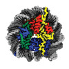
| Entry | Database: PDB / ID: 8cww | ||||||
|---|---|---|---|---|---|---|---|
| Title | Structure of S. cerevisiae Hop1 CBR bound to a nucleosome | ||||||
 Components Components |
| ||||||
 Keywords Keywords | DNA BINDING PROTEIN/DNA /  meiosis / recombination / chromosome axis / nucleosome / PHD / winged helix / DNA BINDING PROTEIN-DNA complex meiosis / recombination / chromosome axis / nucleosome / PHD / winged helix / DNA BINDING PROTEIN-DNA complex | ||||||
| Function / homology | DNA / DNA (> 10) / DNA (> 100) Function and homology information Function and homology information | ||||||
| Biological species |  Saccharomyces cerevisiae (brewer's yeast)Xenopus laevis (African clawed frog) Saccharomyces cerevisiae (brewer's yeast)Xenopus laevis (African clawed frog) Escherichia coli (E. coli) Escherichia coli (E. coli) | ||||||
| Method | ELECTRON MICROSCOPY / single particle reconstruction / cryo EM / Resolution: 2.74 Å | ||||||
 Authors Authors | Gu, Y. / Ur, S.N. / Milano, C.R. / Tromer, E.C. / Vale-Silva, L.A. / Hochwagen, A. / Corbett, K.D. | ||||||
| Funding support |  United States, 1items United States, 1items
| ||||||
 Citation Citation | Journal: EMBO J / Year: 2024 Title: Chromatin binding by HORMAD proteins regulates meiotic recombination initiation. Authors: Carolyn R Milano / Sarah N Ur / Yajie Gu / Jessie Zhang / Rachal Allison / George Brown / Matthew J Neale / Eelco C Tromer / Kevin D Corbett / Andreas Hochwagen /   Abstract: The meiotic chromosome axis coordinates chromosome organization and interhomolog recombination in meiotic prophase and is essential for fertility. In S. cerevisiae, the HORMAD protein Hop1 mediates ...The meiotic chromosome axis coordinates chromosome organization and interhomolog recombination in meiotic prophase and is essential for fertility. In S. cerevisiae, the HORMAD protein Hop1 mediates the enrichment of axis proteins at nucleosome-rich islands through a central chromatin-binding region (CBR). Here, we use cryoelectron microscopy to show that the Hop1 CBR directly recognizes bent nucleosomal DNA through a composite interface in its PHD and winged helix-turn-helix domains. Targeted disruption of the Hop1 CBR-nucleosome interface causes a localized reduction of axis protein binding and meiotic DNA double-strand breaks (DSBs) in axis islands and leads to defects in chromosome synapsis. Synthetic effects with mutants of the Hop1 regulator Pch2 suggest that nucleosome binding delays a conformational switch in Hop1 from a DSB-promoting, Pch2-inaccessible state to a DSB-inactive, Pch2-accessible state to regulate the extent of meiotic DSB formation. Phylogenetic analyses of meiotic HORMADs reveal an ancient origin of the CBR, suggesting that the mechanisms we uncover are broadly conserved. | ||||||
| History |
|
- Structure visualization
| Structure viewer | Molecule: MolmilJmol/JSmol |
|---|
- Downloads & links
-Download
| PDBx/mmCIF format | 8cww.cif.gz | 408.7 KB | Display | PDBx/mmCIF format |
|---|---|---|---|---|
| PDB format | pdb8cww.ent.gz | 258.3 KB | Display | PDB format |
| PDBx/mmJSON format | 8cww.json.gz | Tree view | PDBx/mmJSON format | |
| Others |  Other downloads Other downloads |
-Validation report
| Arichive directory | https://data.pdbj.org/pub/pdb/validation_reports/cw/8cwwftp://data.pdbj.org/pub/pdb/validation_reports/cw/8cww | HTTPS FTP |
|---|
-Related structure data
| Related structure data |  27030MC  7ubaC  8czeC M: map data used to model this data C: citing same article ( |
|---|---|
| Similar structure data |
-Links
 PDBj
PDBj


- Assembly
| Deposited unit | 
|
|---|---|
| 1 |
|
-Components
-Protein , 5 types, 9 molecules PAEBFCGDH
| #1: Protein | Mass: 24239.928 Da / Num. of mol.: 1 Source method: isolated from a genetically manipulated source Details: GenBank:DAA08478.1 Source: (gene. exp.) Saccharomyces cerevisiae (brewer's yeast)Strain: ATCC 204508 / S288c / Gene: HOP1, YIL072W / Production host: Escherichia coli (E. coli) | ||||||
|---|---|---|---|---|---|---|---|
| #2: Protein | Mass: 15303.930 Da / Num. of mol.: 2 Source method: isolated from a genetically manipulated source Details: GenBank:CAD89679.1 / Source: (gene. exp.) Xenopus laevis (African clawed frog) / Production host: Escherichia coli (E. coli)#3: Protein | Mass: 11263.231 Da / Num. of mol.: 2 Source method: isolated from a genetically manipulated source Details: GenBank:NP_001087926.1 / Source: (gene. exp.) Xenopus laevis (African clawed frog) / Production host: Escherichia coli (E. coli)#4: Protein | Mass: 13978.241 Da / Num. of mol.: 2 Source method: isolated from a genetically manipulated source Details: GenBank:CAD89676.1 / Source: (gene. exp.) Xenopus laevis (African clawed frog) / Production host: Escherichia coli (E. coli)#5: Protein | Mass: 13524.752 Da / Num. of mol.: 2 Source method: isolated from a genetically manipulated source Details: GenBank:CAD89678.1 / Source: (gene. exp.) Xenopus laevis (African clawed frog) / Production host: Escherichia coli (E. coli) |
-Widom 601 DNA (146- ... , 2 types, 2 molecules IJ
| #6: DNA chain | Mass: 45274.840 Da / Num. of mol.: 1 / Source method: obtained synthetically / Source: (synth.) Escherichia coli (E. coli) |
|---|---|
| #7: DNA chain | Mass: 44856.570 Da / Num. of mol.: 1 / Source method: obtained synthetically / Source: (synth.) Escherichia coli (E. coli) |
-Non-polymers , 1 types, 2 molecules 
| #8: Chemical |  Mass: 65.409 Da / Num. of mol.: 2 / Source method: obtained synthetically / Formula: Zn / Feature type: SUBJECT OF INVESTIGATION Mass: 65.409 Da / Num. of mol.: 2 / Source method: obtained synthetically / Formula: Zn / Feature type: SUBJECT OF INVESTIGATION |
|---|
-Details
| Has ligand of interest | Y |
|---|
-Experimental details
-Experiment
| Experiment | Method: ELECTRON MICROSCOPY |
|---|---|
| EM experiment | Aggregation state: PARTICLE / 3D reconstruction method: single particle reconstruction |
- Sample preparation
| Component | Name: Complex of Hop1 CBR domain bound to nucleosome / Type: COMPLEX / Entity ID: #1-#7 / Source: MULTIPLE SOURCES | ||||||||||||
|---|---|---|---|---|---|---|---|---|---|---|---|---|---|
| Molecular weight | Value: 0.223 MDa / Experimental value: NO | ||||||||||||
| Source (natural) |
| ||||||||||||
| Source (recombinant) | Organism: Escherichia coli (E. coli) | ||||||||||||
| Buffer solution | pH: 7.5 / Details: 20mM Tris 7.5, 50mM NaCl, 1mM DTT, 1mM EDTA | ||||||||||||
| Specimen | Conc.: 0.8 mg/ml / Embedding applied: NO / Shadowing applied: NO / Staining applied: NO / Vitrification applied: YES | ||||||||||||
| Specimen support | Grid material: COPPER / Grid mesh size: 300 divisions/in. / Grid type: Quantifoil R1.2/1.3 | ||||||||||||
| Vitrification | Cryogen name: ETHANE |
- Electron microscopy imaging
| Experimental equipment |  Model: Titan Krios / Image courtesy: FEI Company |
|---|---|
| Microscopy | Model: FEI TITAN KRIOS |
| Electron gun | Electron source: FIELD EMISSION GUN / Accelerating voltage: 300 kV / Illumination mode: FLOOD BEAM |
| Electron lens | Mode: BRIGHT FIELDBright-field microscopy / Nominal magnification: 130000 X / Nominal defocus max: 2500 nm / Nominal defocus min: 500 nm |
| Image recording | Average exposure time: 10 sec. / Electron dose: 50 e/Å2 / Detector mode: COUNTING / Film or detector model: GATAN K2 QUANTUM (4k x 4k) |
- Processing
| Software |
| ||||||||||||||||||||||||||||||||
|---|---|---|---|---|---|---|---|---|---|---|---|---|---|---|---|---|---|---|---|---|---|---|---|---|---|---|---|---|---|---|---|---|---|
| EM software |
| ||||||||||||||||||||||||||||||||
| CTF correction | Type: PHASE FLIPPING AND AMPLITUDE CORRECTION | ||||||||||||||||||||||||||||||||
| Symmetry | Point symmetry: C1 (asymmetric) | ||||||||||||||||||||||||||||||||
| 3D reconstruction | Resolution: 2.74 Å / Resolution method: FSC 0.143 CUT-OFF / Num. of particles: 139629 / Symmetry type: POINT | ||||||||||||||||||||||||||||||||
| Atomic model building | Protocol: AB INITIO MODEL / Space: REAL | ||||||||||||||||||||||||||||||||
| Refinement | Cross valid method: NONE Stereochemistry target values: GeoStd + Monomer Library + CDL v1.2 | ||||||||||||||||||||||||||||||||
| Displacement parameters | Biso mean: 110.4 Å2 | ||||||||||||||||||||||||||||||||
| Refine LS restraints |
|
