 Movie
Movie Controller

[English] 日本語
 Yorodumi
- PDB-8bdz: Hepatitis B virus core antigen (HBc) with the insertion of four e... -
+ Open data

- Basic information
| Entry | Database: PDB / ID: 8bdz | ||||||
|---|---|---|---|---|---|---|---|
| Title | Hepatitis B virus core antigen (HBc) with the insertion of four external domains of the influenza A M2 protein (HBc/4M2e) with T=4 topology | ||||||
 Components Components | Core protein,Matrix protein 2,External core antigen | ||||||
 Keywords Keywords |  VIRUS LIKE PARTICLE / HBc / M2e / VLP VIRUS LIKE PARTICLE / HBc / M2e / VLP | ||||||
| Function / homology |  Function and homology information Function and homology informationsuppression by virus of host autophagy / microtubule-dependent intracellular transport of viral material towards nucleus / T=4 icosahedral viral capsid / proton transmembrane transporter activity / : / viral penetration into host nucleus / protein complex oligomerization / monoatomic ion channel activity / host cell cytoplasm / virus-mediated perturbation of host defense response ...suppression by virus of host autophagy / microtubule-dependent intracellular transport of viral material towards nucleus / T=4 icosahedral viral capsid / proton transmembrane transporter activity / : / viral penetration into host nucleus / protein complex oligomerization / monoatomic ion channel activity / host cell cytoplasm / virus-mediated perturbation of host defense response / symbiont entry into host cell / host cell nucleus / host cell plasma membrane / virion membrane / structural molecule activity / DNA binding / RNA binding / extracellular region / membraneSimilarity search - Function | ||||||
| Biological species |  Hepatitis B virus adw/991 Hepatitis B virus adw/991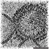 Influenza A virus Influenza A virusHepatitis B virus genotype D subtype ayw | ||||||
| Method | ELECTRON MICROSCOPY / single particle reconstruction / cryo EM / Resolution: 3.13 Å | ||||||
 Authors Authors | Egorov, V.V. / Shvetsov, A.V. / Pichkur, E.B. / Shaldzhyan, A.A. / Zabrodskaya, Y.A. / Vinogradova, D.S. / Nekrasov, P.A. / Gorshkov, A.N. / Garmay, Y.P. / Kovaleva, A.A. ...Egorov, V.V. / Shvetsov, A.V. / Pichkur, E.B. / Shaldzhyan, A.A. / Zabrodskaya, Y.A. / Vinogradova, D.S. / Nekrasov, P.A. / Gorshkov, A.N. / Garmay, Y.P. / Kovaleva, A.A. / Stepanova, L.A. / Tsybalova, L.M. / Shtam, T.A. / Myasnikov, A.G. / Konevega, A.L. | ||||||
| Funding support |  Russian Federation, 1items Russian Federation, 1items
| ||||||
 Citation Citation | Journal: Biophys Chem / Year: 2023 Title: Inside and outside of virus-like particles HBc and HBc/4M2e: A comprehensive study of the structure. Authors: V V Egorov / A V Shvetsov / E B Pichkur / A A Shaldzhyan / Ya A Zabrodskaya / D S Vinogradova / P A Nekrasov / A N Gorshkov / Yu P Garmay / A A Kovaleva / L A Stepanova / L M Tsybalova / T A ...Authors: V V Egorov / A V Shvetsov / E B Pichkur / A A Shaldzhyan / Ya A Zabrodskaya / D S Vinogradova / P A Nekrasov / A N Gorshkov / Yu P Garmay / A A Kovaleva / L A Stepanova / L M Tsybalova / T A Shtam / A G Myasnikov / A L Konevega / Abstract: Hepatitis B virus core antigen (HBc) with the insertion of four external domains of the influenza A M2 protein (HBc/4M2e) form virus-like particles whose structure was studied using a combination of ...Hepatitis B virus core antigen (HBc) with the insertion of four external domains of the influenza A M2 protein (HBc/4M2e) form virus-like particles whose structure was studied using a combination of molecular modeling and cryo-electron microscopy (cryo-EM). It was also shown that self-assembling of the particles occurs inside bacterial cells, but despite the big inner volume of the core shell particle, purified HBc/4M2e contain an insignificant amount of bacterial proteins. It was shown that a fragment of the M2e corresponding to 4M2e insertion is prone to formation of amyloid-like fibrils. However, as the part of the immunodominant loop, M2e insertion does not show a tendency to intermolecular interaction. A full-atomic HBc-4M2e model with the resolution of about 3 Å (3.13 Å for particles of Т = 4 symmetry, 3.7 Å for particles of Т = 3 symmetry) was obtained by molecular modeling methods based on cryo-EM data. | ||||||
| History |
|
- Structure visualization
| Structure viewer | Molecule: MolmilJmol/JSmol |
|---|
- Downloads & links
-Download
| PDBx/mmCIF format | 8bdz.cif.gz | 88.8 KB | Display | PDBx/mmCIF format |
|---|---|---|---|---|
| PDB format | pdb8bdz.ent.gz | 65 KB | Display | PDB format |
| PDBx/mmJSON format | 8bdz.json.gz | Tree view | PDBx/mmJSON format | |
| Others |  Other downloads Other downloads |
-Validation report
| Arichive directory | https://data.pdbj.org/pub/pdb/validation_reports/bd/8bdzftp://data.pdbj.org/pub/pdb/validation_reports/bd/8bdz | HTTPS FTP |
|---|
-Related structure data
| Related structure data |  15995MC 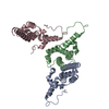 8berC M: map data used to model this data C: citing same article ( |
|---|---|
| Similar structure data |

-Links
 PDBj
PDBj

- Assembly
| Deposited unit | 
|
|---|---|
| 1 |
|
-Components
| #1: Protein | Mass: 30860.533 Da / Num. of mol.: 3 Source method: isolated from a genetically manipulated source Source: (gene. exp.) Hepatitis B virus adw/991, (gene. exp.) Influenza A virus (A/Malaya/302/1954(H1N1)), (gene. exp.) Hepatitis B virus genotype D subtype ayw (isolate France/Tiollais/1979)Gene: prec/C, M, M2 / Strain: A/Malaysia:Malaya/302/1954 H1N1 / Production host:  Escherichia coli (E. coli) Escherichia coli (E. coli)References: UniProt: Q9E0P3, UniProt: A4K144, UniProt: P0C573 |
|---|
-Experimental details
-Experiment
| Experiment | Method: ELECTRON MICROSCOPY |
|---|---|
| EM experiment | Aggregation state: PARTICLE / 3D reconstruction method: single particle reconstruction |
- Sample preparation
| Component | Name: Hepatitis B virus core antigen (HBc) with the insertion of four external domains of the influenza A M2 protein (T=4) Type: COMPLEX / Entity ID: all / Source: RECOMBINANT |
|---|---|
| Molecular weight | Value: 30.84 kDa/nm / Experimental value: NO |
| Source (natural) | Organism:  Hepatitis B virus Hepatitis B virus |
| Source (recombinant) | Organism: Escherichia col (E. coli) |
| Buffer solution | pH: 7.5 |
| Specimen | Embedding applied: NO / Shadowing applied: NO / Staining applied: NO / Vitrification applied: YES |
| Specimen support | Grid material: COPPER / Grid mesh size: 300 divisions/in. / Grid type: Quantifoil R1.2/1.3 |
| Vitrification | Instrument: FEI VITROBOT MARK IV / Cryogen name: ETHANE / Humidity: 100 % / Chamber temperature: 277 K |
- Electron microscopy imaging
| Experimental equipment |  Model: Titan Krios / Image courtesy: FEI Company |
|---|---|
| Microscopy | Model: FEI TITAN KRIOS |
| Electron gun | Electron source: FIELD EMISSION GUN / Accelerating voltage: 300 kV / Illumination mode: SPOT SCAN |
| Electron lens | Mode: BRIGHT FIELDBright-field microscopy / Nominal defocus max: 3000 nm / Nominal defocus min: 1200 nm / Cs: 0.05 mm / C2 aperture diameter: 100 µm / Alignment procedure: ZEMLIN TABLEAU |
| Specimen holder | Cryogen: NITROGEN / Specimen holder model: FEI TITAN KRIOS AUTOGRID HOLDER |
| Image recording | Electron dose: 60 e/Å2 / Detector mode: INTEGRATING / Film or detector model: FEI FALCON II (4k x 4k) |
- Processing
| Software |
| ||||||||||||||||||||||||||||
|---|---|---|---|---|---|---|---|---|---|---|---|---|---|---|---|---|---|---|---|---|---|---|---|---|---|---|---|---|---|
| EM software |
| ||||||||||||||||||||||||||||
| CTF correction | Type: PHASE FLIPPING AND AMPLITUDE CORRECTION | ||||||||||||||||||||||||||||
| Particle selection | Num. of particles selected: 63404 | ||||||||||||||||||||||||||||
| Symmetry | Point symmetry: I (icosahedral) | ||||||||||||||||||||||||||||
| 3D reconstruction | Resolution: 3.13 Å / Resolution method: FSC 0.143 CUT-OFF / Num. of particles: 13146 / Symmetry type: POINT | ||||||||||||||||||||||||||||
| Refinement | Cross valid method: NONE Stereochemistry target values: GeoStd + Monomer Library + CDL v1.2 | ||||||||||||||||||||||||||||
| Displacement parameters | Biso mean: 68.95 Å2 | ||||||||||||||||||||||||||||
| Refine LS restraints |
|
