 Movie
Movie Controller


+ Open data

- Basic information
| Entry | Database: PDB / ID: 8a6u | ||||||
|---|---|---|---|---|---|---|---|
| Title | PcIDS1 in complex with Mg2+ | ||||||
 Components Components | Isoprenyl diphosphate synthase | ||||||
 Keywords Keywords |  BIOSYNTHETIC PROTEIN / Insects / Biosynthesis / Terpenes / Metal regulation / Catalysis BIOSYNTHETIC PROTEIN / Insects / Biosynthesis / Terpenes / Metal regulation / Catalysis | ||||||
| Function / homology |  Function and homology information Function and homology informationpheromone biosynthetic process / dimethylallyltranstransferase activity / isoprenoid biosynthetic process / metal ion bindingSimilarity search - Function | ||||||
| Biological species |  Phaedon cochleariae (mustard beetle) Phaedon cochleariae (mustard beetle) | ||||||
| Method | X-RAY DIFFRACTION / SYNCHROTRON / MOLECULAR REPLACEMENT / Resolution: 1.65 Å | ||||||
 Authors Authors | Ecker, F. / Boland, W. / Groll, M. | ||||||
| Funding support |  Germany, 1items Germany, 1items
| ||||||
 Citation Citation | Journal: Nat.Chem. / Year: 2023 Title: Metal-dependent enzyme symmetry guides the biosynthetic flux of terpene precursors. Authors: Ecker, F. / Vattekkatte, A. / Boland, W. / Groll, M. | ||||||
| History |
|
- Structure visualization
| Structure viewer | Molecule: MolmilJmol/JSmol |
|---|
- Downloads & links
-Download
| PDBx/mmCIF format | 8a6u.cif.gz | 298.4 KB | Display | PDBx/mmCIF format |
|---|---|---|---|---|
| PDB format | pdb8a6u.ent.gz | 241.4 KB | Display | PDB format |
| PDBx/mmJSON format | 8a6u.json.gz | Tree view | PDBx/mmJSON format | |
| Others |  Other downloads Other downloads |
-Validation report
| Arichive directory | https://data.pdbj.org/pub/pdb/validation_reports/a6/8a6uftp://data.pdbj.org/pub/pdb/validation_reports/a6/8a6u | HTTPS FTP |
|---|
-Related structure data
| Related structure data | 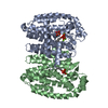 8a6vC 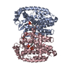 8a6zC 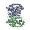 8a70C 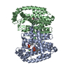 8a73C 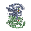 8a74C 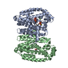 8a78C 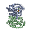 8a7aC 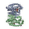 8a7bC 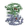 8a7cC 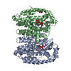 8a7jC 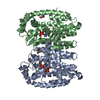 8a7kC  8a7lC 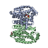 8a7rC 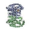 8a7uC 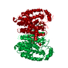 1ubxS S: Starting model for refinement C: citing same article ( |
|---|---|
| Similar structure data |
-Links
 PDBj
PDBj
- Assembly
| Deposited unit | 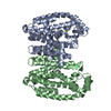
| ||||||||
|---|---|---|---|---|---|---|---|---|---|
| 1 |
| ||||||||
| Unit cell |
|
-Components
| #1: Protein | Mass: 39978.965 Da / Num. of mol.: 2 Source method: isolated from a genetically manipulated source Source: (gene. exp.) Phaedon cochleariae (mustard beetle) / Production host:  Escherichia coli (E. coli) / References: UniProt: M1JS91, dimethylallyltranstransferase Escherichia coli (E. coli) / References: UniProt: M1JS91, dimethylallyltranstransferase#2: Chemical |   Mass: 24.305 Da / Num. of mol.: 2 / Source method: obtained synthetically / Formula: Mg / Feature type: SUBJECT OF INVESTIGATION Mass: 24.305 Da / Num. of mol.: 2 / Source method: obtained synthetically / Formula: Mg / Feature type: SUBJECT OF INVESTIGATION#3: Water | ChemComp-HOH / | Water Mass: 18.015 Da / Num. of mol.: 594 / Source method: isolated from a natural source / Formula: H2O Mass: 18.015 Da / Num. of mol.: 594 / Source method: isolated from a natural source / Formula: H2OHas ligand of interest | Y | |
|---|
-Experimental details
-Experiment
| Experiment | Method: X-RAY DIFFRACTION / Number of used crystals: 1 |
|---|
- Sample preparation
| Crystal | Density Matthews: 2.54 Å3/Da / Density % sol: 51.63 % |
|---|---|
| Crystal grow | Temperature: 293 K / Method: vapor diffusion, sitting drop / pH: 8.5 / Details: 0.1 M TRIS, 0.2 M MgCl2, 20% PEG 8000 |
-Data collection
| Diffraction | Mean temperature: 100 K / Serial crystal experiment: N |
|---|---|
| Diffraction source | Source: SYNCHROTRON / Site: SLS  / Beamline: X06SA / Wavelength: 1 Å / Beamline: X06SA / Wavelength: 1 Å |
| Detector | Type: DECTRIS EIGER X 16M / Detector: PIXEL / Date: Jul 15, 2017 |
| Radiation | Protocol: SINGLE WAVELENGTH / Monochromatic (M) / Laue (L): M / Scattering type: x-ray |
| Radiation wavelength | Wavelength: 1 Å / Relative weight: 1 |
| Reflection | Resolution: 1.65→30 Å / Num. obs: 94381 / % possible obs: 98 % / Redundancy: 3.1 % / Rmerge(I) obs: 0.035 / Net I/σ(I): 16.4 |
| Reflection shell | Resolution: 1.65→1.75 Å / Rmerge(I) obs: 0.488 / Mean I/σ(I) obs: 2.4 / Num. unique obs: 15316 |
- Processing
| Software |
| |||||||||||||||||||||||||||||||||||||||||||||||||||||||||||||||||
|---|---|---|---|---|---|---|---|---|---|---|---|---|---|---|---|---|---|---|---|---|---|---|---|---|---|---|---|---|---|---|---|---|---|---|---|---|---|---|---|---|---|---|---|---|---|---|---|---|---|---|---|---|---|---|---|---|---|---|---|---|---|---|---|---|---|---|
| Refinement | Method to determine structure: MOLECULAR REPLACEMENT Starting model: 1UBX Resolution: 1.65→15 Å / Cor.coef. Fo:Fc: 0.976 / Cor.coef. Fo:Fc free: 0.965 / SU B: 6.381 / SU ML: 0.089 / Cross valid method: THROUGHOUT / σ(F): 0 / ESU R: 0.103 / ESU R Free: 0.084 / Stereochemistry target values: MAXIMUM LIKELIHOOD Details: HYDROGENS HAVE BEEN ADDED IN THE RIDING POSITIONS U VALUES : REFINED INDIVIDUALLY
| |||||||||||||||||||||||||||||||||||||||||||||||||||||||||||||||||
| Solvent computation | Ion probe radii: 0.8 Å / Shrinkage radii: 0.8 Å / VDW probe radii: 1.2 Å / Solvent model: MASK | |||||||||||||||||||||||||||||||||||||||||||||||||||||||||||||||||
| Displacement parameters | Biso max: 105.16 Å2 / Biso mean: 36.621 Å2 / Biso min: 18.54 Å2
| |||||||||||||||||||||||||||||||||||||||||||||||||||||||||||||||||
| Refinement step | Cycle: final / Resolution: 1.65→15 Å
| |||||||||||||||||||||||||||||||||||||||||||||||||||||||||||||||||
| Refine LS restraints |
| |||||||||||||||||||||||||||||||||||||||||||||||||||||||||||||||||
| LS refinement shell | Resolution: 1.65→1.692 Å / Rfactor Rfree error: 0 / Total num. of bins used: 20
|
