 Movie
Movie Controller

[English] 日本語
 Yorodumi
- PDB-7ziq: BK Polyomavirus VP1 in complex with 6'-Sialyllactose glycomacromo... -
+ Open data

- Basic information
| Entry | Database: PDB / ID: 7ziq | ||||||
|---|---|---|---|---|---|---|---|
| Title | BK Polyomavirus VP1 in complex with 6'-Sialyllactose glycomacromolecules (aromatic linker) | ||||||
 Components Components | Capsid protein VP1 | ||||||
 Keywords Keywords | VIRAL PROTEIN / Complex / Sialic acid / Capsid protein | ||||||
| Function / homology |  Function and homology information Function and homology informationT=7 icosahedral viral capsid / virion attachment to host cell / structural molecule activity Similarity search - Function | ||||||
| Biological species |  Betapolyomavirus hominis Betapolyomavirus hominis | ||||||
| Method | X-RAY DIFFRACTION / SYNCHROTRON / MOLECULAR REPLACEMENT / Resolution: 1.9 Å | ||||||
 Authors Authors | Freytag, J. / Mueller, J.C. / Stehle, T. | ||||||
| Funding support |  Germany, 1items Germany, 1items
| ||||||
 Citation Citation | Journal: Biomacromolecules / Year: 2022 Title: Synthesis of Homo- and Heteromultivalent Fucosylated and Sialylated Oligosaccharide Conjugates via Preactivated N -Methyloxyamine Precision Macromolecules and Their Binding to Polyomavirus Capsid Proteins. Authors: Konietzny, P.B. / Freytag, J. / Feldhof, M.I. / Muller, J.C. / Ohl, D. / Stehle, T. / Hartmann, L. | ||||||
| History |
|
- Structure visualization
| Structure viewer | Molecule: MolmilJmol/JSmol |
|---|
- Downloads & links
-Download
| PDBx/mmCIF format | 7ziq.cif.gz | 278.1 KB | Display | PDBx/mmCIF format |
|---|---|---|---|---|
| PDB format | pdb7ziq.ent.gz | Display | PDB format | |
| PDBx/mmJSON format | 7ziq.json.gz | Tree view | PDBx/mmJSON format | |
| Others |  Other downloads Other downloads |
-Validation report
| Arichive directory | https://data.pdbj.org/pub/pdb/validation_reports/zi/7ziqftp://data.pdbj.org/pub/pdb/validation_reports/zi/7ziq | HTTPS FTP |
|---|
-Related structure data
| Related structure data | 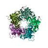 7zilC 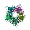 7zimC 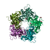 7zinC 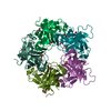 7zioC 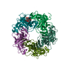 7zipC  4mj1S S: Starting model for refinement C: citing same article ( |
|---|---|
| Similar structure data |
-Links
 PDBj
PDBj


- Assembly
| Deposited unit | 
| ||||||||
|---|---|---|---|---|---|---|---|---|---|
| 1 |
| ||||||||
| Unit cell |
|
-Components
-Protein , 1 types, 5 molecules AAABBBCCCDDDEEE
| #1: Protein | Mass: 30261.039 Da / Num. of mol.: 5 Source method: isolated from a genetically manipulated source Source: (gene. exp.) Betapolyomavirus hominis / Gene: VP1, vp1, BK1035_00004, BK1055_00004 / Production host:  Escherichia coli BL21(DE3) (bacteria) / References: UniProt: Q65613 Escherichia coli BL21(DE3) (bacteria) / References: UniProt: Q65613 |
|---|
-Sugars , 2 types, 3 molecules 
| #2: Polysaccharide | N-acetyl-alpha-neuraminic acid-(2-6)-beta-D-galactopyranose-(1-4)-beta-D-glucopyranose / Mass: 633.552 Da / Num. of mol.: 1 Source method: isolated from a genetically manipulated source |
|---|---|
| #4: Sugar | Sialic acid Type: D-saccharide, alpha linking / Mass: 309.270 Da / Num. of mol.: 2 / Source method: obtained synthetically / Formula: C11H19NO9 / Feature type: SUBJECT OF INVESTIGATION Type: D-saccharide, alpha linking / Mass: 309.270 Da / Num. of mol.: 2 / Source method: obtained synthetically / Formula: C11H19NO9 / Feature type: SUBJECT OF INVESTIGATION |
-Non-polymers , 3 types, 753 molecules 


| #3: Chemical | ChemComp-EDO / Ethylene glycol Mass: 62.068 Da / Num. of mol.: 4 Mass: 62.068 Da / Num. of mol.: 4Source method: isolated from a genetically manipulated source Formula: C2H6O2 / Feature type: SUBJECT OF INVESTIGATION #5: Chemical | ChemComp-PEG / | Diethylene glycol Mass: 106.120 Da / Num. of mol.: 1 / Source method: obtained synthetically / Formula: C4H10O3 Mass: 106.120 Da / Num. of mol.: 1 / Source method: obtained synthetically / Formula: C4H10O3#6: Water | ChemComp-HOH / | WaterMass: 18.015 Da / Num. of mol.: 748 / Source method: isolated from a natural source / Formula: H2O |
|---|
-Details
| Has ligand of interest | Y |
|---|
-Experimental details
-Experiment
| Experiment | Method: X-RAY DIFFRACTION / Number of used crystals: 1 |
|---|
- Sample preparation
| Crystal | Density Matthews: 2.28 Å3/Da / Density % sol: 46.12 % |
|---|---|
| Crystal grow | Temperature: 293 K / Method: vapor diffusion, hanging drop Details: 0.1 M HEPES pH 7.5, 0.25 M LiCl, 18% (w/v) PEG 3350 |
-Data collection
| Diffraction | Mean temperature: 93 K / Serial crystal experiment: N |
|---|---|
| Diffraction source | Source: SYNCHROTRON / Site: SLS  / Beamline: X06DA / Wavelength: 0.99 Å / Beamline: X06DA / Wavelength: 0.99 Å |
| Detector | Type: DECTRIS PILATUS 2M-F / Detector: PIXEL / Date: Feb 5, 2022 |
| Radiation | Protocol: SINGLE WAVELENGTH / Monochromatic (M) / Laue (L): M / Scattering type: x-ray |
| Radiation wavelength | Wavelength: 0.99 Å / Relative weight: 1 |
| Reflection | Resolution: 1.9→48.5 Å / Num. obs: 110045 / % possible obs: 99.9 % / Redundancy: 13.42 % / CC1/2: 0.99 / Rrim(I) all: 0.19 / Net I/σ(I): 12.84 |
| Reflection shell | Resolution: 1.9→2.01 Å / Num. unique obs: 17476 / CC1/2: 0.61 / Rrim(I) all: 1.94 |
- Processing
| Software |
| |||||||||||||||||||||||||||||||||||||||||||||||||||||||||||||||||||||||||||||||||||||||||||||||||||||||||||||||||||||||||||||||||||||||||||||||||||||||||||
|---|---|---|---|---|---|---|---|---|---|---|---|---|---|---|---|---|---|---|---|---|---|---|---|---|---|---|---|---|---|---|---|---|---|---|---|---|---|---|---|---|---|---|---|---|---|---|---|---|---|---|---|---|---|---|---|---|---|---|---|---|---|---|---|---|---|---|---|---|---|---|---|---|---|---|---|---|---|---|---|---|---|---|---|---|---|---|---|---|---|---|---|---|---|---|---|---|---|---|---|---|---|---|---|---|---|---|---|---|---|---|---|---|---|---|---|---|---|---|---|---|---|---|---|---|---|---|---|---|---|---|---|---|---|---|---|---|---|---|---|---|---|---|---|---|---|---|---|---|---|---|---|---|---|---|---|---|
| Refinement | Method to determine structure: MOLECULAR REPLACEMENT Starting model: 4MJ1 Resolution: 1.9→48.5 Å / Cor.coef. Fo:Fc: 0.964 / Cor.coef. Fo:Fc free: 0.945 / Cross valid method: FREE R-VALUE / ESU R: 0.148 / ESU R Free: 0.138 Details: Hydrogens have been added in their riding positions
| |||||||||||||||||||||||||||||||||||||||||||||||||||||||||||||||||||||||||||||||||||||||||||||||||||||||||||||||||||||||||||||||||||||||||||||||||||||||||||
| Solvent computation | Ion probe radii: 0.8 Å / Shrinkage radii: 0.8 Å / VDW probe radii: 1.2 Å / Solvent model: MASK BULK SOLVENT | |||||||||||||||||||||||||||||||||||||||||||||||||||||||||||||||||||||||||||||||||||||||||||||||||||||||||||||||||||||||||||||||||||||||||||||||||||||||||||
| Displacement parameters | Biso mean: 30.528 Å2
| |||||||||||||||||||||||||||||||||||||||||||||||||||||||||||||||||||||||||||||||||||||||||||||||||||||||||||||||||||||||||||||||||||||||||||||||||||||||||||
| Refinement step | Cycle: LAST / Resolution: 1.9→48.5 Å
| |||||||||||||||||||||||||||||||||||||||||||||||||||||||||||||||||||||||||||||||||||||||||||||||||||||||||||||||||||||||||||||||||||||||||||||||||||||||||||
| Refine LS restraints |
| |||||||||||||||||||||||||||||||||||||||||||||||||||||||||||||||||||||||||||||||||||||||||||||||||||||||||||||||||||||||||||||||||||||||||||||||||||||||||||
| LS refinement shell |
|
