 Movie
Movie Controller

[English] 日本語
 Yorodumi
- PDB-7z6r: Psychrobacter arcticus ATPPRT (HisGZ) R56A mutant bound to ATP an... -
+ Open data

- Basic information
| Entry | Database: PDB / ID: 7z6r | ||||||
|---|---|---|---|---|---|---|---|
| Title | Psychrobacter arcticus ATPPRT (HisGZ) R56A mutant bound to ATP and PRPP | ||||||
 Components Components |
| ||||||
 Keywords Keywords |  TRANSFERASE / phosphoribosyltransferase / ATPPRT / PRPP / HisGZ / histidine / biosynthesis / allosteric TRANSFERASE / phosphoribosyltransferase / ATPPRT / PRPP / HisGZ / histidine / biosynthesis / allosteric | ||||||
| Function / homology |  Function and homology informationATP phosphoribosyltransferase / ATP phosphoribosyltransferase activity / L-histidine biosynthetic process / ATP binding / cytoplasm Function and homology informationATP phosphoribosyltransferase / ATP phosphoribosyltransferase activity / L-histidine biosynthetic process / ATP binding / cytoplasmSimilarity search - Function | ||||||
| Biological species |  Psychrobacter arcticus (bacteria) Psychrobacter arcticus (bacteria) | ||||||
| Method | X-RAY DIFFRACTION / SYNCHROTRON / MOLECULAR REPLACEMENT / molecular replacement / Resolution: 2.55 Å | ||||||
 Authors Authors | Alphey, M.S. / Fisher, G. / da Silva, R.G. | ||||||
| Funding support |  United Kingdom, 1items United Kingdom, 1items
| ||||||
 Citation Citation | Journal: Nat Commun / Year: 2022 Title: Allosteric rescue of catalytically impaired ATP phosphoribosyltransferase variants links protein dynamics to active-site electrostatic preorganisation. Authors: Fisher, G. / Corbella, M. / Alphey, M.S. / Nicholson, J. / Read, B.J. / Kamerlin, S.C.L. / da Silva, R.G. | ||||||
| History |
|
- Structure visualization
| Structure viewer | Molecule: MolmilJmol/JSmol |
|---|
- Downloads & links
-Download
| PDBx/mmCIF format | 7z6r.cif.gz | 432.5 KB | Display | PDBx/mmCIF format |
|---|---|---|---|---|
| PDB format | pdb7z6r.ent.gz | 354.7 KB | Display | PDB format |
| PDBx/mmJSON format | 7z6r.json.gz | Tree view | PDBx/mmJSON format | |
| Others |  Other downloads Other downloads |
-Validation report
| Arichive directory | https://data.pdbj.org/pub/pdb/validation_reports/z6/7z6rftp://data.pdbj.org/pub/pdb/validation_reports/z6/7z6r | HTTPS FTP |
|---|
-Related structure data
| Related structure data |  7z8uC  6fu2S S: Starting model for refinement C: citing same article ( |
|---|---|
| Similar structure data |
-Links
 PDBj
PDBj

- Assembly
| Deposited unit | 
| ||||||||||||||||||||||||||||||||||||||||||||||||||||||||||||||||||||
|---|---|---|---|---|---|---|---|---|---|---|---|---|---|---|---|---|---|---|---|---|---|---|---|---|---|---|---|---|---|---|---|---|---|---|---|---|---|---|---|---|---|---|---|---|---|---|---|---|---|---|---|---|---|---|---|---|---|---|---|---|---|---|---|---|---|---|---|---|---|
| 1 |
| ||||||||||||||||||||||||||||||||||||||||||||||||||||||||||||||||||||
| Unit cell |
| ||||||||||||||||||||||||||||||||||||||||||||||||||||||||||||||||||||
| Noncrystallographic symmetry (NCS) | NCS domain:
NCS domain segments: Component-ID: 0 / Refine code: 0
NCS ensembles :
|
-Components
| #1: Protein | Mass: 43129.039 Da / Num. of mol.: 2 Source method: isolated from a genetically manipulated source Source: (gene. exp.) Psychrobacter arcticus (bacteria) / Gene: hisZ, Psyc_0676 / Production host: Escherichia coli BL21(DE3) (bacteria) / References: UniProt: Q4FTX3#2: Protein | / ATP-PRT / ATP-PRTaseMass: 25154.850 Da / Num. of mol.: 2 / Mutation: R56A Source method: isolated from a genetically manipulated source Source: (gene. exp.) Psychrobacter arcticus (bacteria) / Strain: DSM 17307 / VKM B-2377 / 273-4 / Gene: hisG, Psyc_1903 / Production host: Escherichia coli BL21(DE3) (bacteria) / Variant (production host): C43 / References: UniProt: Q4FQF7, ATP phosphoribosyltransferase#3: Chemical | Adenosine triphosphate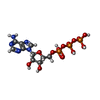  Mass: 507.181 Da / Num. of mol.: 2 / Source method: obtained synthetically / Formula: C10H16N5O13P3 / Feature type: SUBJECT OF INVESTIGATION / Comment: ATP, energy-carrying molecule*YM Mass: 507.181 Da / Num. of mol.: 2 / Source method: obtained synthetically / Formula: C10H16N5O13P3 / Feature type: SUBJECT OF INVESTIGATION / Comment: ATP, energy-carrying molecule*YM#4: Sugar | Phosphoribosyl pyrophosphate  Type: D-saccharide / Mass: 390.070 Da / Num. of mol.: 2 / Source method: obtained synthetically / Formula: C5H13O14P3 / Feature type: SUBJECT OF INVESTIGATION Type: D-saccharide / Mass: 390.070 Da / Num. of mol.: 2 / Source method: obtained synthetically / Formula: C5H13O14P3 / Feature type: SUBJECT OF INVESTIGATION#5: Water | ChemComp-HOH / | Water Mass: 18.015 Da / Num. of mol.: 16 / Source method: isolated from a natural source / Formula: H2O Mass: 18.015 Da / Num. of mol.: 16 / Source method: isolated from a natural source / Formula: H2OHas ligand of interest | Y | |
|---|
-Experimental details
-Experiment
| Experiment | Method: X-RAY DIFFRACTION / Number of used crystals: 1 |
|---|
- Sample preparation
| Crystal | Density Matthews: 2.48 Å3/Da / Density % sol: 50.44 % |
|---|---|
| Crystal grow | Temperature: 277 K / Method: vapor diffusion, hanging drop / pH: 8.5 Details: 8mg mL-1 protein (in 20mM Tris pH7, 50mM KCl, 10mM MgCl2, 2mM DTT, 0.5mM histidine) mixed in 1:1 ratio with precipitant solution (12% PEG3350, 100mM bicine pH8.5, 150mM SrCl2, 150mM KBr, 2% 1,6-hexanediol) |
-Data collection
| Diffraction | Mean temperature: 100 K / Serial crystal experiment: N | ||||||||||||||||||||||||
|---|---|---|---|---|---|---|---|---|---|---|---|---|---|---|---|---|---|---|---|---|---|---|---|---|---|
| Diffraction source | Source: SYNCHROTRON / Site: Diamond / Beamline: I03 / Wavelength: 0.97628 Å | ||||||||||||||||||||||||
| Detector | Type: DECTRIS EIGER2 XE 16M / Detector: PIXEL / Date: May 11, 2021 | ||||||||||||||||||||||||
| Radiation | Protocol: SINGLE WAVELENGTH / Monochromatic (M) / Laue (L): M / Scattering type: x-ray | ||||||||||||||||||||||||
| Radiation wavelength | Wavelength: 0.97628 Å / Relative weight: 1 | ||||||||||||||||||||||||
| Reflection | Resolution: 2.55→56.97 Å / Num. obs: 43754 / % possible obs: 99.9 % / Redundancy: 6.5 % / Biso Wilson estimate: 53.1 Å2 / Rmerge(I) obs: 0.172 / Rpim(I) all: 0.073 / Rrim(I) all: 0.187 / Net I/σ(I): 10.9 | ||||||||||||||||||||||||
| Reflection shell | Diffraction-ID: 1
|
-Phasing
| Phasing | Method: molecular replacement | ||||||
|---|---|---|---|---|---|---|---|
| Phasing MR | R rigid body: 0.523
|
- Processing
| Software |
| |||||||||||||||||||||||||||||||||||||||||||||||||||||||||||||||||||||||||||||||||||||||||||||||||||||||||||||||||||||||||||||
|---|---|---|---|---|---|---|---|---|---|---|---|---|---|---|---|---|---|---|---|---|---|---|---|---|---|---|---|---|---|---|---|---|---|---|---|---|---|---|---|---|---|---|---|---|---|---|---|---|---|---|---|---|---|---|---|---|---|---|---|---|---|---|---|---|---|---|---|---|---|---|---|---|---|---|---|---|---|---|---|---|---|---|---|---|---|---|---|---|---|---|---|---|---|---|---|---|---|---|---|---|---|---|---|---|---|---|---|---|---|---|---|---|---|---|---|---|---|---|---|---|---|---|---|---|---|---|
| Refinement | Method to determine structure: MOLECULAR REPLACEMENT Starting model: 6FU2 Resolution: 2.55→56.97 Å / Cor.coef. Fo:Fc: 0.943 / Cor.coef. Fo:Fc free: 0.92 / WRfactor Rfree: 0.2508 / WRfactor Rwork: 0.2098 / FOM work R set: 0.6855 / SU B: 39.307 / SU ML: 0.359 / SU R Cruickshank DPI: 0.645 / SU Rfree: 0.3229 / Cross valid method: THROUGHOUT / σ(F): 0 / ESU R: 0.645 / ESU R Free: 0.323 / Stereochemistry target values: MAXIMUM LIKELIHOOD Details: HYDROGENS HAVE BEEN ADDED IN THE RIDING POSITIONS U VALUES : WITH TLS ADDED
| |||||||||||||||||||||||||||||||||||||||||||||||||||||||||||||||||||||||||||||||||||||||||||||||||||||||||||||||||||||||||||||
| Solvent computation | Ion probe radii: 0.7 Å / Shrinkage radii: 0.7 Å / VDW probe radii: 1 Å / Solvent model: MASK | |||||||||||||||||||||||||||||||||||||||||||||||||||||||||||||||||||||||||||||||||||||||||||||||||||||||||||||||||||||||||||||
| Displacement parameters | Biso max: 144.86 Å2 / Biso mean: 68.237 Å2 / Biso min: 36.89 Å2
| |||||||||||||||||||||||||||||||||||||||||||||||||||||||||||||||||||||||||||||||||||||||||||||||||||||||||||||||||||||||||||||
| Refinement step | Cycle: final / Resolution: 2.55→56.97 Å
| |||||||||||||||||||||||||||||||||||||||||||||||||||||||||||||||||||||||||||||||||||||||||||||||||||||||||||||||||||||||||||||
| Refine LS restraints |
| |||||||||||||||||||||||||||||||||||||||||||||||||||||||||||||||||||||||||||||||||||||||||||||||||||||||||||||||||||||||||||||
| Refine LS restraints NCS | Refine-ID: X-RAY DIFFRACTION / Type: interatomic distance / Weight position: 0.05
| |||||||||||||||||||||||||||||||||||||||||||||||||||||||||||||||||||||||||||||||||||||||||||||||||||||||||||||||||||||||||||||
| LS refinement shell | Resolution: 2.55→2.611 Å / Rfactor Rfree error: 0
| |||||||||||||||||||||||||||||||||||||||||||||||||||||||||||||||||||||||||||||||||||||||||||||||||||||||||||||||||||||||||||||
| Refinement TLS params. | Method: refined / Refine-ID: X-RAY DIFFRACTION
| |||||||||||||||||||||||||||||||||||||||||||||||||||||||||||||||||||||||||||||||||||||||||||||||||||||||||||||||||||||||||||||
| Refinement TLS group |
|
