 Movie
Movie Controller

[English] 日本語
 Yorodumi
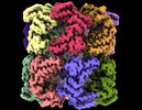
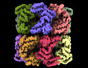
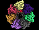
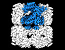
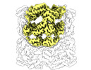
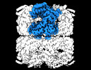
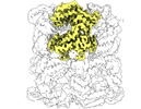
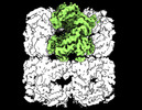
- PDB-7xor: Cryo-EM structure of occupied ring subunit 3 (OR3) of GroEL from ... -
+ Open data

- Basic information
| Entry | Database: PDB / ID: 7xor | ||||||||||||||||||||||||
|---|---|---|---|---|---|---|---|---|---|---|---|---|---|---|---|---|---|---|---|---|---|---|---|---|---|
| Title | Cryo-EM structure of occupied ring subunit 3 (OR3) of GroEL from GroEL-UGT1A double occupied ring complex | ||||||||||||||||||||||||
 Components Components | Chaperonin GroEL | ||||||||||||||||||||||||
 Keywords Keywords |  CHAPERONE / cryogenic electron microscopy / single-particle analysis / molecular motion / structure-function relationship / focus classification / separating heterogeneity / GroEL / unfolded protein CHAPERONE / cryogenic electron microscopy / single-particle analysis / molecular motion / structure-function relationship / focus classification / separating heterogeneity / GroEL / unfolded protein | ||||||||||||||||||||||||
| Function / homology |  Function and homology information Function and homology informationGroEL-GroES complex / chaperonin ATPase / virion assembly / chaperone cofactor-dependent protein refolding / isomerase activity / ATP-dependent protein folding chaperone / response to radiation / unfolded protein binding / protein folding / response to heat ...GroEL-GroES complex / chaperonin ATPase / virion assembly / chaperone cofactor-dependent protein refolding / isomerase activity / ATP-dependent protein folding chaperone / response to radiation / unfolded protein binding / protein folding / response to heat / protein refolding / magnesium ion binding / ATP hydrolysis activity / ATP binding / membrane / identical protein binding / cytosolSimilarity search - Function | ||||||||||||||||||||||||
| Biological species |  Escherichia coli (E. coli) Escherichia coli (E. coli) | ||||||||||||||||||||||||
| Method | ELECTRON MICROSCOPY / single particle reconstruction / cryo EM / Resolution: 3.3 Å | ||||||||||||||||||||||||
 Authors Authors | Stapleton, K. / Takagi, J. / Mizohata, E. | ||||||||||||||||||||||||
| Funding support |  Japan, 7items Japan, 7items
| ||||||||||||||||||||||||
 Citation Citation | Journal: To Be Published Title: Unmasking GroEL: Structure, dynamics, and substrate binding revealed by single-particle cryo-EM Authors: Stapleton, K.M. / Mizobata, T. / Miyazaki, N. / Takatsuji, T. / Kato, T. / Iwasaki, K. / Standley, D.M. / Kawamura, T. / Nakane, T. / Takagi, J. / Mizohata, E. | ||||||||||||||||||||||||
| History |
|
- Structure visualization
| Structure viewer | Molecule: MolmilJmol/JSmol |
|---|
- Downloads & links
-Download
| PDBx/mmCIF format | 7xor.cif.gz | 94.2 KB | Display | PDBx/mmCIF format |
|---|---|---|---|---|
| PDB format | pdb7xor.ent.gz | 72.5 KB | Display | PDB format |
| PDBx/mmJSON format | 7xor.json.gz | Tree view | PDBx/mmJSON format | |
| Others |  Other downloads Other downloads |
-Validation report
| Arichive directory | https://data.pdbj.org/pub/pdb/validation_reports/xo/7xorftp://data.pdbj.org/pub/pdb/validation_reports/xo/7xor | HTTPS FTP |
|---|
-Related structure data
| Related structure data |  33357MC  7xojC  7xokC  7xolC  7xomC 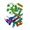 7xonC 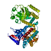 7xooC  7xopC 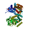 7xoqC 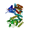 7xosC M: map data used to model this data C: citing same article ( |
|---|---|
| Similar structure data |

-Links
 PDBj
PDBj
- Assembly
| Deposited unit | 
|
|---|---|
| 1 |
|
-Components
| #1: Protein | Mass: 57260.504 Da / Num. of mol.: 1 Source method: isolated from a genetically manipulated source Source: (gene. exp.) Escherichia coli (E. coli) / Strain: K12 / Gene: groEL, groL, mopA, b4143, JW4103 / Production host: Escherichia coli (E. coli) / References: UniProt: P0A6F5, chaperonin ATPase |
|---|
-Experimental details
-Experiment
| Experiment | Method: ELECTRON MICROSCOPY |
|---|---|
| EM experiment | Aggregation state: PARTICLE / 3D reconstruction method: single particle reconstruction |
- Sample preparation
| Component |
| ||||||||||||||||||||||||||||
|---|---|---|---|---|---|---|---|---|---|---|---|---|---|---|---|---|---|---|---|---|---|---|---|---|---|---|---|---|---|
| Molecular weight |
| ||||||||||||||||||||||||||||
| Source (natural) |
| ||||||||||||||||||||||||||||
| Source (recombinant) |
| ||||||||||||||||||||||||||||
| Buffer solution | pH: 7.5 Details: Sample containing GorEL-UGT1A was made fresh and used without undergoing any freeze-thaw cycles to avoid degradation in the solution. The sample was in a buffer solution of 150mM NaCl, 20mM Tris-HCl at pH 7.5 | ||||||||||||||||||||||||||||
| Buffer component |
| ||||||||||||||||||||||||||||
| Specimen | Conc.: 33 mg/ml / Embedding applied: NO / Shadowing applied: NO / Staining applied: NO / Vitrification applied: YES | ||||||||||||||||||||||||||||
| Specimen support | Grid material: COPPER / Grid mesh size: 300 divisions/in. / Grid type: Quantifoil | ||||||||||||||||||||||||||||
| Vitrification | Instrument: FEI VITROBOT MARK IV / Cryogen name: ETHANE / Humidity: 100 % / Chamber temperature: 277.15 K Details: 3 ul of 33 mg/ml GroEL-UGT1A was placed on Holey carbon Quanitifoil copper grids (300 mesh size R1.2/1.3) and blotted for 3 seconds (blot force =1) |

- Electron microscopy imaging
| Experimental equipment |  Model: Titan Krios / Image courtesy: FEI Company |
|---|---|
| Microscopy | Model: FEI TITAN KRIOS |
| Electron gun | Electron source: FIELD EMISSION GUN / Accelerating voltage: 300 kV / Illumination mode: FLOOD BEAM |
| Electron lens | Mode: BRIGHT FIELDBright-field microscopy / Nominal defocus max: 2000 nm / Nominal defocus min: 1200 nm / C2 aperture diameter: 50 µm |
| Specimen holder | Cryogen: NITROGEN / Specimen holder model: FEI TITAN KRIOS AUTOGRID HOLDER |
| Image recording | Electron dose: 40 e/Å2 / Detector mode: COUNTING / Film or detector model: FEI FALCON III (4k x 4k) / Num. of grids imaged: 1 / Num. of real images: 2951 Details: Using a Titan KRIOS TEM operated at 300 kV, 2,951 movies were collected using the Falcon III DED in Counting mode at a magnification of 75000x corresponding to a pixel size of 0.87 Pixel/A ...Details: Using a Titan KRIOS TEM operated at 300 kV, 2,951 movies were collected using the Falcon III DED in Counting mode at a magnification of 75000x corresponding to a pixel size of 0.87 Pixel/A at the specimen level. All 2,951 movies were imported into the RELION pipeline and prepared for single-particle analysis |
| EM imaging optics | Energyfilter slit width: 20 eV |
- Processing
| Software | Name: PHENIX / Version: 1.19.2_4158: / Classification: refinement | |||||||||||||||||||||||||||||||||||||||||||||
|---|---|---|---|---|---|---|---|---|---|---|---|---|---|---|---|---|---|---|---|---|---|---|---|---|---|---|---|---|---|---|---|---|---|---|---|---|---|---|---|---|---|---|---|---|---|---|
| EM software |
| |||||||||||||||||||||||||||||||||||||||||||||
| CTF correction | Details: Using RELION's own implementation of CTFFIND4.1 --Box 512 --ResMin 30 --ResMax 2 --dFMin 5000 --dFMax 50000 --FStep 250 --dAst 100 Type: PHASE FLIPPING ONLY | |||||||||||||||||||||||||||||||||||||||||||||
| Particle selection | Num. of particles selected: 1289236 Details: Parameters for auto picking (LoG) single particles from motion-corrected micrographs with estimated CTF parameters resulted in 1,289,236 particles picked (an average of 437 particles per ...Details: Parameters for auto picking (LoG) single particles from motion-corrected micrographs with estimated CTF parameters resulted in 1,289,236 particles picked (an average of 437 particles per micrograph). All particle images were then extracted with a box size of 300 pixels and a spherical mask diameter of 240 A for 2D classification. | |||||||||||||||||||||||||||||||||||||||||||||
| Symmetry | Point symmetry: C1 (asymmetric) | |||||||||||||||||||||||||||||||||||||||||||||
| 3D reconstruction | Resolution: 3.3 Å / Resolution method: FSC 0.143 CUT-OFF / Num. of particles: 60993 / Algorithm: FOURIER SPACE / Num. of class averages: 1 / Symmetry type: POINT | |||||||||||||||||||||||||||||||||||||||||||||
| Atomic model building | Protocol: RIGID BODY FIT / Space: REAL | |||||||||||||||||||||||||||||||||||||||||||||
| Refine LS restraints |
|
