 Movie
Movie Controller

[English] 日本語
 Yorodumi
- PDB-7wij: Cryo-EM structure of prenyltransferase domain of Macrophoma phase... -
+ Open data

- Basic information
| Entry | Database: PDB / ID: 7wij | ||||||
|---|---|---|---|---|---|---|---|
| Title | Cryo-EM structure of prenyltransferase domain of Macrophoma phaseolina macrophomene synthase | ||||||
 Components Components | Geranylgeranyl diphosphate synthase | ||||||
 Keywords Keywords |  TRANSFERASE / Macrophoma phaseolina / Macrophomene Synthase / prenyltransferase TRANSFERASE / Macrophoma phaseolina / Macrophomene Synthase / prenyltransferase | ||||||
| Function / homology | geranylgeranyl diphosphate synthase / Polyprenyl synthases signature 1. / isoprenoid biosynthetic process / Polyprenyl synthetase, conserved site / Polyprenyl synthetase / Polyprenyl synthetase / Isoprenoid synthase domain superfamily / Geranylgeranyl diphosphate synthase Function and homology information Function and homology information | ||||||
| Biological species |  Macrophomina phaseolina MS6 (fungus) Macrophomina phaseolina MS6 (fungus) | ||||||
| Method | ELECTRON MICROSCOPY / single particle reconstruction / cryo EM / Resolution: 3.17 Å | ||||||
 Authors Authors | Mori, T. / Adachi, N. / Abe, I. | ||||||
| Funding support |  Japan, 1items Japan, 1items
| ||||||
 Citation Citation | Journal: Nature / Year: 2022 Title: Discovery of non-squalene triterpenes. Authors: Hui Tao / Lukas Lauterbach / Guangkai Bian / Rong Chen / Anwei Hou / Takahiro Mori / Shu Cheng / Ben Hu / Li Lu / Xin Mu / Min Li / Naruhiko Adachi / Masato Kawasaki / Toshio Moriya / ...Authors: Hui Tao / Lukas Lauterbach / Guangkai Bian / Rong Chen / Anwei Hou / Takahiro Mori / Shu Cheng / Ben Hu / Li Lu / Xin Mu / Min Li / Naruhiko Adachi / Masato Kawasaki / Toshio Moriya / Toshiya Senda / Xinghuan Wang / Zixin Deng / Ikuro Abe / Jeroen S Dickschat / Tiangang Liu /   Abstract: All known triterpenes are generated by triterpene synthases (TrTSs) from squalene or oxidosqualene. This approach is fundamentally different from the biosynthesis of short-chain (C-C) terpenes that ...All known triterpenes are generated by triterpene synthases (TrTSs) from squalene or oxidosqualene. This approach is fundamentally different from the biosynthesis of short-chain (C-C) terpenes that are formed from polyisoprenyl diphosphates. In this study, two fungal chimeric class I TrTSs, Talaromyces verruculosus talaropentaene synthase (TvTS) and Macrophomina phaseolina macrophomene synthase (MpMS), were characterized. Both enzymes use dimethylallyl diphosphate and isopentenyl diphosphate or hexaprenyl diphosphate as substrates, representing the first examples, to our knowledge, of non-squalene-dependent triterpene biosynthesis. The cyclization mechanisms of TvTS and MpMS and the absolute configurations of their products were investigated in isotopic labelling experiments. Structural analyses of the terpene cyclase domain of TvTS and full-length MpMS provide detailed insights into their catalytic mechanisms. An AlphaFold2-based screening platform was developed to mine a third TrTS, Colletotrichum gloeosporioides colleterpenol synthase (CgCS). Our findings identify a new enzymatic mechanism for the biosynthesis of triterpenes and enhance understanding of terpene biosynthesis in nature. | ||||||
| History |
|
- Structure visualization
| Structure viewer | Molecule: MolmilJmol/JSmol |
|---|
- Downloads & links
-Download
| PDBx/mmCIF format | 7wij.cif.gz | 311.6 KB | Display | PDBx/mmCIF format |
|---|---|---|---|---|
| PDB format | pdb7wij.ent.gz | 233.8 KB | Display | PDB format |
| PDBx/mmJSON format | 7wij.json.gz | Tree view | PDBx/mmJSON format | |
| Others |  Other downloads Other downloads |
-Validation report
| Arichive directory | https://data.pdbj.org/pub/pdb/validation_reports/wi/7wijftp://data.pdbj.org/pub/pdb/validation_reports/wi/7wij | HTTPS FTP |
|---|
-Related structure data
| Related structure data | 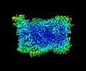 32531MC  7vtaC  7vtbC M: map data used to model this data C: citing same article ( |
|---|---|
| Similar structure data |

-Links
 PDBj
PDBj
- Assembly
| Deposited unit | 
|
|---|---|
| 1 |
|
-Components
| #1: Protein | Mass: 80442.664 Da / Num. of mol.: 6 Source method: isolated from a genetically manipulated source Source: (gene. exp.) Macrophomina phaseolina MS6 (fungus) / Strain: MS6 / Gene: MPH_02178 / Production host:  Escherichia coli (E. coli) Escherichia coli (E. coli)References: UniProt: K2SUY0, geranylgeranyl diphosphate synthase |
|---|
-Experimental details
-Experiment
| Experiment | Method: ELECTRON MICROSCOPY |
|---|---|
| EM experiment | Aggregation state: PARTICLE / 3D reconstruction method: single particle reconstruction |
- Sample preparation
| Component | Name: Macrophoma phaseolina macrophomene synthase / Type: ORGANELLE OR CELLULAR COMPONENT / Entity ID: all / Source: RECOMBINANT | ||||||||||||||||||||
|---|---|---|---|---|---|---|---|---|---|---|---|---|---|---|---|---|---|---|---|---|---|
| Source (natural) | Organism: Macrophomina phaseolina | ||||||||||||||||||||
| Source (recombinant) | Organism: Escherichia coli (E. coli) | ||||||||||||||||||||
| Buffer solution | pH: 7.5 | ||||||||||||||||||||
| Buffer component |
| ||||||||||||||||||||
| Specimen | Conc.: 2 mg/ml / Embedding applied: NO / Shadowing applied: NO / Staining applied: NO / Vitrification applied: YES / Details: This sample was mono-disperse | ||||||||||||||||||||
| Specimen support | Details: This grid was washed with acetone prior to use / Grid material: COPPER / Grid mesh size: 300 divisions/in. / Grid type: Quantifoil R1.2/1.3 | ||||||||||||||||||||
| Vitrification | Instrument: FEI VITROBOT MARK IV / Cryogen name: ETHANE / Humidity: 100 % / Chamber temperature: 291 K / Details: Blotting time was 20 second (blot fource 0) |
- Electron microscopy imaging
| Experimental equipment |  Model: Talos Arctica / Image courtesy: FEI Company |
|---|---|
| Microscopy | Model: FEI TALOS ARCTICA |
| Electron gun | Electron source: FIELD EMISSION GUN / Accelerating voltage: 200 kV / Illumination mode: FLOOD BEAM |
| Electron lens | Mode: BRIGHT FIELDBright-field microscopy / Nominal magnification: 120000 X / Nominal defocus max: 2500 nm / Nominal defocus min: 1000 nm / Cs: 2.7 mm / C2 aperture diameter: 50 µm |
| Specimen holder | Cryogen: NITROGEN |
| Image recording | Electron dose: 50 e/Å2 / Detector mode: COUNTING / Film or detector model: FEI FALCON III (4k x 4k) / Num. of grids imaged: 1 / Num. of real images: 1888 |
- Processing
| Software |
| ||||||||||||||||||||||||||||||||||||||||
|---|---|---|---|---|---|---|---|---|---|---|---|---|---|---|---|---|---|---|---|---|---|---|---|---|---|---|---|---|---|---|---|---|---|---|---|---|---|---|---|---|---|
| EM software |
| ||||||||||||||||||||||||||||||||||||||||
| CTF correction | Type: PHASE FLIPPING AND AMPLITUDE CORRECTION | ||||||||||||||||||||||||||||||||||||||||
| Symmetry | Point symmetry: D3 (2x3 fold dihedral) | ||||||||||||||||||||||||||||||||||||||||
| 3D reconstruction | Resolution: 3.17 Å / Resolution method: FSC 0.143 CUT-OFF / Num. of particles: 132926 / Algorithm: FOURIER SPACE / Symmetry type: POINT | ||||||||||||||||||||||||||||||||||||||||
| Atomic model building | Protocol: OTHER / Space: REAL | ||||||||||||||||||||||||||||||||||||||||
| Refinement | Cross valid method: NONE Stereochemistry target values: GeoStd + Monomer Library + CDL v1.2 | ||||||||||||||||||||||||||||||||||||||||
| Displacement parameters | Biso mean: 35.95 Å2 | ||||||||||||||||||||||||||||||||||||||||
| Refine LS restraints |
|
