 Movie
Movie Controller


+ Open data

- Basic information
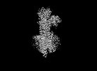
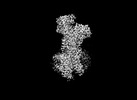
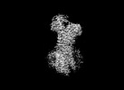
| Entry | Database: PDB / ID: 7w7w | |||||||||||||||
|---|---|---|---|---|---|---|---|---|---|---|---|---|---|---|---|---|
| Title | E2 Pi of SERCA2b | |||||||||||||||
 Components Components | Sarcoplasmic/endoplasmic reticulum calcium ATPase 2 | |||||||||||||||
 Keywords Keywords | METAL TRANSPORT /  calcium calcium | |||||||||||||||
| Function / homology |  Function and homology information Function and homology informationlongitudinal sarcoplasmic reticulum / ER-nucleus signaling pathway / P-type calcium transporter activity involved in regulation of cardiac muscle cell membrane potential / regulation of calcium ion-dependent exocytosis of neurotransmitter / calcium ion transport from cytosol to endoplasmic reticulum / positive regulation of endoplasmic reticulum calcium ion concentration / calcium ion-transporting ATPase complex / T-tubule organization / regulation of cardiac muscle cell action potential involved in regulation of contraction / regulation of cardiac muscle cell membrane potential ...longitudinal sarcoplasmic reticulum / ER-nucleus signaling pathway / P-type calcium transporter activity involved in regulation of cardiac muscle cell membrane potential / regulation of calcium ion-dependent exocytosis of neurotransmitter / calcium ion transport from cytosol to endoplasmic reticulum / positive regulation of endoplasmic reticulum calcium ion concentration / calcium ion-transporting ATPase complex / T-tubule organization / regulation of cardiac muscle cell action potential involved in regulation of contraction / regulation of cardiac muscle cell membrane potential / ribbon synapse / platelet dense tubular network membrane / calcium ion import into sarcoplasmic reticulum / Pre-NOTCH Processing in Golgi / P-type Ca2+ transporter / sarcoplasmic reticulum calcium ion transport / negative regulation of heart contraction / P-type calcium transporter activity / regulation of the force of heart contraction / transition between fast and slow fiber / cardiac muscle hypertrophy in response to stress / endoplasmic reticulum calcium ion homeostasis / regulation of cardiac muscle contraction by calcium ion signaling / lncRNA binding / S100 protein binding / Reduction of cytosolic Ca++ levels / relaxation of cardiac muscle / organelle localization by membrane tethering / mitochondrion-endoplasmic reticulum membrane tethering / autophagosome membrane docking / positive regulation of heart rate / positive regulation of cardiac muscle cell apoptotic process / Ion transport by P-type ATPases / autophagosome assembly / regulation of cardiac conduction / epidermis development / Ion homeostasis / sarcoplasmic reticulum membrane / response to endoplasmic reticulum stress / monoatomic ion transmembrane transport / sarcoplasmic reticulum / negative regulation of receptor binding / calcium ion transmembrane transport / intracellular calcium ion homeostasis / neuron cellular homeostasis / cellular response to oxidative stress / transmembrane transporter binding / cell adhesion / calcium ion binding / endoplasmic reticulum membrane / enzyme binding / endoplasmic reticulum / ATP hydrolysis activity / ATP binding / membrane / plasma membraneSimilarity search - Function | |||||||||||||||
| Biological species |  Homo sapiens (human) Homo sapiens (human) | |||||||||||||||
| Method | ELECTRON MICROSCOPY / single particle reconstruction / cryo EM / Resolution: 3.2 Å | |||||||||||||||
 Authors Authors | Zhang, Y. / Watanabe, S. / Tsutsumi, A. / Inaba, K. | |||||||||||||||
| Funding support |  Japan, 4items Japan, 4items
| |||||||||||||||
 Citation Citation | Journal: Cell Rep / Year: 2022 Title: Multiple sub-state structures of SERCA2b reveal conformational overlap at transition steps during the catalytic cycle. Authors: Yuxia Zhang / Chigusa Kobayashi / Xiaohan Cai / Satoshi Watanabe / Akihisa Tsutsumi / Masahide Kikkawa / Yuji Sugita / Kenji Inaba / Abstract: Sarco/endoplasmic reticulum Ca ATPase (SERCA) pumps Ca into the endoplasmic reticulum (ER). Herein, we present cryo-electron microscopy (EM) structures of three intermediates of SERCA2b: Ca-bound ...Sarco/endoplasmic reticulum Ca ATPase (SERCA) pumps Ca into the endoplasmic reticulum (ER). Herein, we present cryo-electron microscopy (EM) structures of three intermediates of SERCA2b: Ca-bound phosphorylated (E1P·2Ca) and Ca-unbound dephosphorylated (E2·Pi) intermediates and another between the E2P and E2·Pi states. Our cryo-EM analysis demonstrates that the E1P·2Ca state exists in low abundance and preferentially transitions to an E2P-like structure by releasing Ca and that the Ca release gate subsequently undergoes stepwise closure during the dephosphorylation processes. Importantly, each intermediate adopts multiple sub-state structures including those like the next one in the catalytic series, indicating conformational overlap at transition steps, as further substantiated by atomistic molecular dynamic simulations of SERCA2b in a lipid bilayer. The present findings provide insight into how enzymes accelerate catalytic cycles. #1: Journal: EMBO J / Year: 2021Title: Cryo-EM analysis provides new mechanistic insight into ATP binding to Ca -ATPase SERCA2b. Authors: Yuxia Zhang / Satoshi Watanabe / Akihisa Tsutsumi / Hiroshi Kadokura / Masahide Kikkawa / Kenji Inaba / Abstract: Sarco/endoplasmic reticulum Ca -ATPase (SERCA) 2b is a ubiquitous SERCA family member that conducts Ca uptake from the cytosol to the ER. Herein, we present a 3.3 Å resolution cryo-electron ...Sarco/endoplasmic reticulum Ca -ATPase (SERCA) 2b is a ubiquitous SERCA family member that conducts Ca uptake from the cytosol to the ER. Herein, we present a 3.3 Å resolution cryo-electron microscopy (cryo-EM) structure of human SERCA2b in the E1·2Ca state, revealing a new conformation for Ca -bound SERCA2b with a much closer arrangement of cytosolic domains than in the previously reported crystal structure of Ca -bound SERCA1a. Multiple conformations generated by 3D classification of cryo-EM maps reflect the intrinsically dynamic nature of the cytosolic domains in this state. Notably, ATP binding residues of SERCA2b in the E1·2Ca state are located at similar positions to those in the E1·2Ca -ATP state; hence, the cryo-EM structure likely represents a preformed state immediately prior to ATP binding. Consistently, a SERCA2b mutant with an interdomain disulfide bridge that locks the closed cytosolic domain arrangement displayed significant autophosphorylation activity in the presence of Ca . We propose a novel mechanism of ATP binding to SERCA2b. | |||||||||||||||
| History |
|
- Structure visualization
| Structure viewer | Molecule: MolmilJmol/JSmol |
|---|
- Downloads & links
-Download
| PDBx/mmCIF format | 7w7w.cif.gz | 181.5 KB | Display | PDBx/mmCIF format |
|---|---|---|---|---|
| PDB format | pdb7w7w.ent.gz | 144.8 KB | Display | PDB format |
| PDBx/mmJSON format | 7w7w.json.gz | Tree view | PDBx/mmJSON format | |
| Others |  Other downloads Other downloads |
-Validation report
| Arichive directory | https://data.pdbj.org/pub/pdb/validation_reports/w7/7w7wftp://data.pdbj.org/pub/pdb/validation_reports/w7/7w7w | HTTPS FTP |
|---|
-Related structure data
| Related structure data |  32350MC  7w7tC  7w7uC  7w7vC M: map data used to model this data C: citing same article ( |
|---|---|
| Similar structure data |
-Links
 PDBj
PDBj


- Assembly
| Deposited unit | 
|
|---|---|
| 1 |
|
-Components
| #1: Protein | Mass: 114869.664 Da / Num. of mol.: 1 Source method: isolated from a genetically manipulated source Source: (gene. exp.) Homo sapiens (human) / Gene: ATP2A2, ATP2B / Production host: Homo sapiens (human) / References: UniProt: P16615, P-type Ca2+ transporter |
|---|---|
| #2: Chemical | ChemComp-ALF / 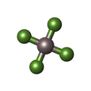  Mass: 102.975 Da / Num. of mol.: 1 / Source method: obtained synthetically / Formula: AlF4 / Feature type: SUBJECT OF INVESTIGATION Mass: 102.975 Da / Num. of mol.: 1 / Source method: obtained synthetically / Formula: AlF4 / Feature type: SUBJECT OF INVESTIGATION |
| #3: Chemical | ChemComp-MG /   Mass: 24.305 Da / Num. of mol.: 1 / Source method: obtained synthetically / Formula: Mg / Feature type: SUBJECT OF INVESTIGATION Mass: 24.305 Da / Num. of mol.: 1 / Source method: obtained synthetically / Formula: Mg / Feature type: SUBJECT OF INVESTIGATION |
| Has ligand of interest | Y |
-Experimental details
-Experiment
| Experiment | Method: ELECTRON MICROSCOPY |
|---|---|
| EM experiment | Aggregation state: PARTICLE / 3D reconstruction method: single particle reconstruction |
- Sample preparation
| Component | Name: SERCA2b with Ca / Type: COMPLEX / Entity ID: #1 / Source: RECOMBINANT |
|---|---|
| Molecular weight | Value: 110 kDa/nm / Experimental value: NO |
| Source (natural) | Organism: Homo sapiens (human) |
| Source (recombinant) | Organism: Homo sapiens (human) |
| Buffer solution | pH: 7 |
| Specimen | Embedding applied: NO / Shadowing applied: NO / Staining applied: NO / Vitrification applied: YES |
| Vitrification | Cryogen name: ETHANE |
- Electron microscopy imaging
| Experimental equipment |  Model: Titan Krios / Image courtesy: FEI Company |
|---|---|
| Microscopy | Model: FEI TITAN KRIOS |
| Electron gun | Electron source: FIELD EMISSION GUN / Accelerating voltage: 300 kV / Illumination mode: OTHER |
| Electron lens | Mode: BRIGHT FIELDBright-field microscopy |
| Image recording | Electron dose: 60 e/Å2 / Film or detector model: GATAN K3 BIOQUANTUM (6k x 4k) |
- Processing
| Software | Name: PHENIX / Version: 1.19.1_4122: / Classification: refinement | ||||||||||||||||||||||||
|---|---|---|---|---|---|---|---|---|---|---|---|---|---|---|---|---|---|---|---|---|---|---|---|---|---|
| CTF correction | Type: NONE | ||||||||||||||||||||||||
| 3D reconstruction | Resolution: 3.2 Å / Resolution method: FSC 0.143 CUT-OFF / Num. of particles: 104446 / Symmetry type: POINT | ||||||||||||||||||||||||
| Refine LS restraints |
|
