 Movie
Movie Controller

[English] 日本語
 Yorodumi
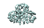
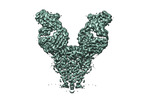
- PDB-7vgr: SARS-CoV-2 M protein dimer (long form) in complex with YN7756_1 Fab -
+ Open data

- Basic information
| Entry | Database: PDB / ID: 7vgr | ||||||
|---|---|---|---|---|---|---|---|
| Title | SARS-CoV-2 M protein dimer (long form) in complex with YN7756_1 Fab | ||||||
 Components Components |
| ||||||
 Keywords Keywords |  VIRAL PROTEIN/IMMUNE SYSTEM / SARS-CoV-2 / M protein / viral structural protein / virus assembly / VIRAL PROTEIN / VIRAL PROTEIN-IMMUNE SYSTEM complex VIRAL PROTEIN/IMMUNE SYSTEM / SARS-CoV-2 / M protein / viral structural protein / virus assembly / VIRAL PROTEIN / VIRAL PROTEIN-IMMUNE SYSTEM complex | ||||||
| Function / homology |  Function and homology information Function and homology informationMaturation of protein M / SARS-CoV-2 modulates autophagy / cytoplasmic capsid assembly / host cell Golgi membrane / CD28 dependent PI3K/Akt signaling / endoplasmic reticulum-Golgi intermediate compartment / SARS-CoV-2 targets host intracellular signalling and regulatory pathways / symbiont-mediated suppression of host cytoplasmic pattern recognition receptor signaling pathway via inhibition of MAVS activity / protein sequestering activity / VEGFR2 mediated vascular permeability ...Maturation of protein M / SARS-CoV-2 modulates autophagy / cytoplasmic capsid assembly / host cell Golgi membrane / CD28 dependent PI3K/Akt signaling / endoplasmic reticulum-Golgi intermediate compartment / SARS-CoV-2 targets host intracellular signalling and regulatory pathways / symbiont-mediated suppression of host cytoplasmic pattern recognition receptor signaling pathway via inhibition of MAVS activity / protein sequestering activity / VEGFR2 mediated vascular permeability / PIP3 activates AKT signaling / TRAF3-dependent IRF activation pathway / Translation of Structural Proteins / Virion Assembly and Release / Induction of Cell-Cell Fusion / structural constituent of virion / Attachment and Entry / viral envelope / SARS-CoV-2 activates/modulates innate and adaptive immune responses / virion membrane / identical protein binding / plasma membraneSimilarity search - Function | ||||||
| Biological species |  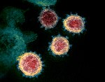 Severe acute respiratory syndrome coronavirus 2 Severe acute respiratory syndrome coronavirus 2 Mus musculus (house mouse) Mus musculus (house mouse) | ||||||
| Method | ELECTRON MICROSCOPY / single particle reconstruction / cryo EM / Resolution: 2.7 Å | ||||||
 Authors Authors | Zhang, Z. / Ohto, U. / Shimizu, T. | ||||||
| Funding support | 1items
| ||||||
 Citation Citation | Journal: Nat Commun / Year: 2022 Title: Structure of SARS-CoV-2 membrane protein essential for virus assembly. Authors: Zhikuan Zhang / Norimichi Nomura / Yukiko Muramoto / Toru Ekimoto / Tomoko Uemura / Kehong Liu / Moeko Yui / Nozomu Kono / Junken Aoki / Mitsunori Ikeguchi / Takeshi Noda / So Iwata / ...Authors: Zhikuan Zhang / Norimichi Nomura / Yukiko Muramoto / Toru Ekimoto / Tomoko Uemura / Kehong Liu / Moeko Yui / Nozomu Kono / Junken Aoki / Mitsunori Ikeguchi / Takeshi Noda / So Iwata / Umeharu Ohto / Toshiyuki Shimizu /  Abstract: The coronavirus membrane protein (M) is the most abundant viral structural protein and plays a central role in virus assembly and morphogenesis. However, the process of M protein-driven virus ...The coronavirus membrane protein (M) is the most abundant viral structural protein and plays a central role in virus assembly and morphogenesis. However, the process of M protein-driven virus assembly are largely unknown. Here, we report the cryo-electron microscopy structure of the SARS-CoV-2 M protein in two different conformations. M protein forms a mushroom-shaped dimer, composed of two transmembrane domain-swapped three-helix bundles and two intravirion domains. M protein further assembles into higher-order oligomers. A highly conserved hinge region is key for conformational changes. The M protein dimer is unexpectedly similar to SARS-CoV-2 ORF3a, a viral ion channel. Moreover, the interaction analyses of M protein with nucleocapsid protein (N) and RNA suggest that the M protein mediates the concerted recruitment of these components through the positively charged intravirion domain. Our data shed light on the M protein-driven virus assembly mechanism and provide a structural basis for therapeutic intervention targeting M protein. | ||||||
| History |
|
- Structure visualization
| Structure viewer | Molecule: MolmilJmol/JSmol |
|---|
- Downloads & links
-Download
| PDBx/mmCIF format | 7vgr.cif.gz | 271.6 KB | Display | PDBx/mmCIF format |
|---|---|---|---|---|
| PDB format | pdb7vgr.ent.gz | 212.9 KB | Display | PDB format |
| PDBx/mmJSON format | 7vgr.json.gz | Tree view | PDBx/mmJSON format | |
| Others |  Other downloads Other downloads |
-Validation report
| Arichive directory | https://data.pdbj.org/pub/pdb/validation_reports/vg/7vgrftp://data.pdbj.org/pub/pdb/validation_reports/vg/7vgr | HTTPS FTP |
|---|
-Related structure data
| Related structure data |  31977MC 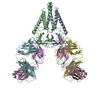 7vgsC M: map data used to model this data C: citing same article ( |
|---|---|
| Similar structure data | |
| EM raw data | EMPIAR-11168 (Title: Structure of SARS-CoV-2 membrane protein / Data size: 4.4 TB / Data #1: M protein (LMNG/CHS) [micrographs - multiframe] Data #2: M protein (LMNG/CHS) + Fab-E [micrographs - multiframe] Data #3: M protein (LMNG/CHS) + Fab-B [micrographs - multiframe]) |
-Links
 PDBj
PDBj


- Assembly
| Deposited unit | 
|
|---|---|
| 1 |
|
-Components
| #1: Antibody | Mass: 24025.412 Da / Num. of mol.: 2 / Source method: isolated from a natural source / Source: (natural) Mus musculus (house mouse)#2: Antibody | Mass: 25114.109 Da / Num. of mol.: 2 / Source method: isolated from a natural source / Source: (natural) Mus musculus (house mouse)#3: Protein | / M / E1 glycoprotein / Matrix glycoprotein / Membrane glycoproteinMass: 28257.822 Da / Num. of mol.: 2 Source method: isolated from a genetically manipulated source Source: (gene. exp.) Severe acute respiratory syndrome coronavirus 2Production host:  Homo sapiens (human) / References: UniProt: P0DTC5 Homo sapiens (human) / References: UniProt: P0DTC5 |
|---|
-Experimental details
-Experiment
| Experiment | Method: ELECTRON MICROSCOPY |
|---|---|
| EM experiment | Aggregation state: PARTICLE / 3D reconstruction method: single particle reconstruction |
- Sample preparation
| Component |
| ||||||||||||||||||||||||
|---|---|---|---|---|---|---|---|---|---|---|---|---|---|---|---|---|---|---|---|---|---|---|---|---|---|
| Molecular weight | Experimental value: NO | ||||||||||||||||||||||||
| Source (natural) |
| ||||||||||||||||||||||||
| Source (recombinant) | Organism: Homo sapiens (human) | ||||||||||||||||||||||||
| Buffer solution | pH: 7.6 | ||||||||||||||||||||||||
| Specimen | Conc.: 2 mg/ml / Embedding applied: NO / Shadowing applied: NO / Staining applied: NO / Vitrification applied: YES | ||||||||||||||||||||||||
| Specimen support | Grid material: COPPER / Grid type: Quantifoil R1.2/1.3 | ||||||||||||||||||||||||
| Vitrification | Cryogen name: ETHANE |
- Electron microscopy imaging
| Experimental equipment |  Model: Titan Krios / Image courtesy: FEI Company |
|---|---|
| Microscopy | Model: TFS KRIOS |
| Electron gun | Electron source: FIELD EMISSION GUN / Accelerating voltage: 300 kV / Illumination mode: OTHER |
| Electron lens | Mode: BRIGHT FIELDBright-field microscopy |
| Image recording | Electron dose: 61.9 e/Å2 / Film or detector model: GATAN K3 (6k x 4k) |
- Processing
| Software | Name: PHENIX / Version: 1.19.2_4158: / Classification: refinement | ||||||||||||||||||||||||
|---|---|---|---|---|---|---|---|---|---|---|---|---|---|---|---|---|---|---|---|---|---|---|---|---|---|
| CTF correction | Type: NONE | ||||||||||||||||||||||||
| 3D reconstruction | Resolution: 2.7 Å / Resolution method: FSC 0.143 CUT-OFF / Num. of particles: 263166 / Symmetry type: POINT | ||||||||||||||||||||||||
| Atomic model building | Protocol: AB INITIO MODEL / Space: REAL | ||||||||||||||||||||||||
| Refine LS restraints |
|
