 Movie
Movie Controller


+ Open data

- Basic information
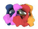
| Entry | Database: PDB / ID: 7ttr | |||||||||
|---|---|---|---|---|---|---|---|---|---|---|
| Title | Skd3_ATPyS_FITC-casein Hexamer, AAA+ only | |||||||||
 Components Components |
| |||||||||
 Keywords Keywords |  CHAPERONE / cryoEM / AAA+ CHAPERONE / cryoEM / AAA+ | |||||||||
| Function / homology |  Function and homology information Function and homology informationgranulocyte differentiation / RIG-I signaling pathway / ATP-dependent protein disaggregase activity / Hydrolases; Acting on acid anhydrides; In phosphorus-containing anhydrides / antiviral innate immune response / mitochondrial intermembrane space / cellular response to heat / ATP hydrolysis activity / mitochondrion / extracellular space ...granulocyte differentiation / RIG-I signaling pathway / ATP-dependent protein disaggregase activity / Hydrolases; Acting on acid anhydrides; In phosphorus-containing anhydrides / antiviral innate immune response / mitochondrial intermembrane space / cellular response to heat / ATP hydrolysis activity / mitochondrion / extracellular space / extracellular region / ATP binding / cytoplasmSimilarity search - Function | |||||||||
| Biological species |  Homo sapiens (human) Homo sapiens (human) Bos taurus (cattle) Bos taurus (cattle) | |||||||||
| Method | ELECTRON MICROSCOPY / single particle reconstruction / cryo EM / Resolution: 2.96 Å | |||||||||
 Authors Authors | Rizo, A.N. | |||||||||
| Funding support |  United States, 2items United States, 2items
| |||||||||
 Citation Citation | Journal: Cell Rep / Year: 2022 Title: Unique structural features govern the activity of a human mitochondrial AAA+ disaggregase, Skd3. Authors: Ryan R Cupo / Alexandrea N Rizo / Gabriel A Braun / Eric Tse / Edward Chuang / Kushol Gupta / Daniel R Southworth / James Shorter / Abstract: The AAA+ protein, Skd3 (human CLPB), solubilizes proteins in the mitochondrial intermembrane space, which is critical for human health. Skd3 variants with defective protein-disaggregase activity ...The AAA+ protein, Skd3 (human CLPB), solubilizes proteins in the mitochondrial intermembrane space, which is critical for human health. Skd3 variants with defective protein-disaggregase activity cause severe congenital neutropenia (SCN) and 3-methylglutaconic aciduria type 7 (MGCA7). How Skd3 disaggregates proteins remains poorly understood. Here, we report a high-resolution structure of a Skd3-substrate complex. Skd3 adopts a spiral hexameric arrangement that engages substrate via pore-loop interactions in the nucleotide-binding domain (NBD). Substrate-bound Skd3 hexamers stack head-to-head via unique, adaptable ankyrin-repeat domain (ANK)-mediated interactions to form dodecamers. Deleting the ANK linker region reduces dodecamerization and disaggregase activity. We elucidate apomorphic features of the Skd3 NBD and C-terminal domain that regulate disaggregase activity. We also define how Skd3 subunits collaborate to disaggregate proteins. Importantly, SCN-linked subunits sharply inhibit disaggregase activity, whereas MGCA7-linked subunits do not. These advances illuminate Skd3 structure and mechanism, explain SCN and MGCA7 inheritance patterns, and suggest therapeutic strategies. | |||||||||
| History |
|
- Structure visualization
| Structure viewer | Molecule: MolmilJmol/JSmol |
|---|
- Downloads & links
-Download
| PDBx/mmCIF format | 7ttr.cif.gz | 365.2 KB | Display | PDBx/mmCIF format |
|---|---|---|---|---|
| PDB format | pdb7ttr.ent.gz | 291.3 KB | Display | PDB format |
| PDBx/mmJSON format | 7ttr.json.gz | Tree view | PDBx/mmJSON format | |
| Others |  Other downloads Other downloads |
-Validation report
| Arichive directory | https://data.pdbj.org/pub/pdb/validation_reports/tt/7ttrftp://data.pdbj.org/pub/pdb/validation_reports/tt/7ttr | HTTPS FTP |
|---|
-Related structure data
| Related structure data |  26121MC  7ttsC C: citing same article ( M: map data used to model this data |
|---|---|
| Similar structure data |
-Links
 PDBj
PDBj


- Assembly
| Deposited unit | 
|
|---|---|
| 1 |
|
-Components
| #1: Protein | Mass: 66115.047 Da / Num. of mol.: 6 Source method: isolated from a genetically manipulated source Source: (gene. exp.) Homo sapiens (human) / Gene: CLPB, HSP78, SKD3 / Production host:  Escherichia coli BL21(DE3) (bacteria) Escherichia coli BL21(DE3) (bacteria)References: UniProt: Q9H078, Hydrolases; Acting on acid anhydrides; In phosphorus-containing anhydrides#2: Protein | | CSN2Mass: 24759.648 Da / Num. of mol.: 1 / Source method: obtained synthetically / Source: (synth.) Bos taurus (cattle) / References: UniProt: T1T0C1#3: Chemical | ChemComp-ADP / | Adenosine diphosphate  Mass: 427.201 Da / Num. of mol.: 1 / Source method: obtained synthetically / Formula: C10H15N5O10P2 / Feature type: SUBJECT OF INVESTIGATION / Comment: ADP, energy-carrying molecule*YM Mass: 427.201 Da / Num. of mol.: 1 / Source method: obtained synthetically / Formula: C10H15N5O10P2 / Feature type: SUBJECT OF INVESTIGATION / Comment: ADP, energy-carrying molecule*YM#4: Chemical | ChemComp-MG /   Mass: 24.305 Da / Num. of mol.: 4 / Source method: obtained synthetically / Formula: Mg / Feature type: SUBJECT OF INVESTIGATION Mass: 24.305 Da / Num. of mol.: 4 / Source method: obtained synthetically / Formula: Mg / Feature type: SUBJECT OF INVESTIGATION#5: Chemical | ChemComp-AGS / 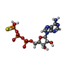  Mass: 523.247 Da / Num. of mol.: 4 / Source method: obtained synthetically / Formula: C10H16N5O12P3S / Feature type: SUBJECT OF INVESTIGATION / Comment: ATP-gamma-S, energy-carrying molecule analogue*YM Mass: 523.247 Da / Num. of mol.: 4 / Source method: obtained synthetically / Formula: C10H16N5O12P3S / Feature type: SUBJECT OF INVESTIGATION / Comment: ATP-gamma-S, energy-carrying molecule analogue*YMHas ligand of interest | Y | Sequence details | The residues that were modeled in the casein were modeled as UNK due to the fact that they could ...The residues that were modeled in the casein were modeled as UNK due to the fact that they could not be identified. The actual sequence of the casein is MKVLILACLV | |
|---|
-Experimental details
-Experiment
| Experiment | Method: ELECTRON MICROSCOPY |
|---|---|
| EM experiment | Aggregation state: PARTICLE / 3D reconstruction method: single particle reconstruction |
- Sample preparation
| Component | Name: Skd3 complex bound to FITC-casein / Type: COMPLEX / Entity ID: #1-#2 / Source: MULTIPLE SOURCES | ||||||||||||
|---|---|---|---|---|---|---|---|---|---|---|---|---|---|
| Molecular weight |
| ||||||||||||
| Buffer solution | pH: 8 | ||||||||||||
| Specimen | Embedding applied: NO / Shadowing applied: NO / Staining applied: NO / Vitrification applied: YES | ||||||||||||
| Specimen support | Grid material: GOLD / Grid mesh size: 300 divisions/in. / Grid type: Quantifoil R1.2/1.3 | ||||||||||||
| Vitrification | Instrument: FEI VITROBOT MARK IV / Cryogen name: ETHANE / Humidity: 100 % |
- Electron microscopy imaging
| Microscopy | Model: FEI TITAN |
|---|---|
| Electron gun | Electron source: FIELD EMISSION GUN / Accelerating voltage: 300 kV / Illumination mode: FLOOD BEAM |
| Electron lens | Mode: BRIGHT FIELDBright-field microscopy / Nominal defocus max: 1500 nm / Nominal defocus min: 800 nm |
| Image recording | Electron dose: 68 e/Å2 / Film or detector model: GATAN K3 (6k x 4k) |
- Processing
| Software | Name: PHENIX / Version: 1.19.1_4122: / Classification: refinement | ||||||||||||||||||||||||
|---|---|---|---|---|---|---|---|---|---|---|---|---|---|---|---|---|---|---|---|---|---|---|---|---|---|
| CTF correction | Type: PHASE FLIPPING AND AMPLITUDE CORRECTION | ||||||||||||||||||||||||
| 3D reconstruction | Resolution: 2.96 Å / Resolution method: FSC 0.143 CUT-OFF / Num. of particles: 358000 / Symmetry type: POINT | ||||||||||||||||||||||||
| Refine LS restraints |
|
