Movie
Movie Controller

[English] 日本語
 Yorodumi
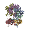
- PDB-7tib: Structure of the yeast clamp loader (Replication Factor C RFC) bo... -
+ Open data

- Basic information
| Entry | Database: PDB / ID: 7tib | |||||||||||||||
|---|---|---|---|---|---|---|---|---|---|---|---|---|---|---|---|---|
| Title | Structure of the yeast clamp loader (Replication Factor C RFC) bound to the open sliding clamp (Proliferating Cell Nuclear Antigen PCNA) and primer-template DNA | |||||||||||||||
 Components Components |
| |||||||||||||||
 Keywords Keywords | REPLICATION/DNA /  sliding clamp / DNA replication / AAA+ / clamp loader / REPLICATION-DNA complex sliding clamp / DNA replication / AAA+ / clamp loader / REPLICATION-DNA complex | |||||||||||||||
| Function / homology |  Function and homology information Function and homology informationDNA clamp unloading / Rad17 RFC-like complex / Mismatch repair (MMR) directed by MSH2:MSH6 (MutSalpha) / meiotic mismatch repair / Processive synthesis on the lagging strand / Removal of the Flap Intermediate / Elg1 RFC-like complex / DNA replication factor C complex / Ctf18 RFC-like complex / DNA clamp loader activity ...DNA clamp unloading / Rad17 RFC-like complex / Mismatch repair (MMR) directed by MSH2:MSH6 (MutSalpha) / meiotic mismatch repair / Processive synthesis on the lagging strand / Removal of the Flap Intermediate / Elg1 RFC-like complex / DNA replication factor C complex / Ctf18 RFC-like complex / DNA clamp loader activity / Polymerase switching / SUMOylation of DNA replication proteins / positive regulation of DNA metabolic process / maintenance of DNA trinucleotide repeats / PCNA complex / establishment of mitotic sister chromatid cohesion / DNA replication checkpoint signaling / Activation of ATR in response to replication stress / lagging strand elongation / postreplication repair / sister chromatid cohesion / silent mating-type cassette heterochromatin formation / mitotic sister chromatid cohesion / leading strand elongation / DNA polymerase processivity factor activity / error-free translesion synthesis / Gap-filling DNA repair synthesis and ligation in TC-NER / subtelomeric heterochromatin formation / mismatch repair / translesion synthesis / positive regulation of DNA repair / DNA damage checkpoint signaling / replication fork / positive regulation of DNA replication / nucleotide-excision repair / DNA-templated DNA replication / mitotic cell cycle / chromosome, telomeric region / cell division / DNA repair / ATP hydrolysis activity / DNA binding / ATP binding / identical protein binding / nucleus / cytosolSimilarity search - Function | |||||||||||||||
| Biological species |  Saccharomyces cerevisiae (brewer's yeast) Saccharomyces cerevisiae (brewer's yeast)synthetic construct (others) | |||||||||||||||
| Method | ELECTRON MICROSCOPY / single particle reconstruction / cryo EM / Resolution: 3.4 Å | |||||||||||||||
 Authors Authors | Gaubitz, C. / Liu, X. / Pajak, J. / Stone, N. / Hayes, J. / Demo, G. / Kelch, B.A. | |||||||||||||||
| Funding support |  United States, 4items United States, 4items
| |||||||||||||||
 Citation Citation | Journal: Elife / Year: 2022 Title: Cryo-EM structures reveal high-resolution mechanism of a DNA polymerase sliding clamp loader. Authors: Christl Gaubitz / Xingchen Liu / Joshua Pajak / Nicholas P Stone / Janelle A Hayes / Gabriel Demo / Brian A Kelch /  Abstract: Sliding clamps are ring-shaped protein complexes that are integral to the DNA replication machinery of all life. Sliding clamps are opened and installed onto DNA by clamp loader AAA+ ATPase complexes. ...Sliding clamps are ring-shaped protein complexes that are integral to the DNA replication machinery of all life. Sliding clamps are opened and installed onto DNA by clamp loader AAA+ ATPase complexes. However, how a clamp loader opens and closes the sliding clamp around DNA is still unknown. Here, we describe structures of the clamp loader Replication Factor C (RFC) bound to its cognate sliding clamp Proliferating Cell Nuclear Antigen (PCNA) en route to successful loading. RFC first binds to PCNA in a dynamic, closed conformation that blocks both ATPase activity and DNA binding. RFC then opens the PCNA ring through a large-scale 'crab-claw' expansion of both RFC and PCNA that explains how RFC prefers initial binding of PCNA over DNA. Next, the open RFC:PCNA complex binds DNA and interrogates the primer-template junction using a surprising base-flipping mechanism. Our structures indicate that initial PCNA opening and subsequent closure around DNA do not require ATP hydrolysis, but are driven by binding energy. ATP hydrolysis, which is necessary for RFC release, is triggered by interactions with both PCNA and DNA, explaining RFC's switch-like ATPase activity. Our work reveals how a AAA+ machine undergoes dramatic conformational changes for achieving binding preference and substrate remodeling. | |||||||||||||||
| History |
|
- Structure visualization
| Movie |
Movie viewer |
|---|---|
| Structure viewer | Molecule: MolmilJmol/JSmol |
- Downloads & links
-Download
| PDBx/mmCIF format | 7tib.cif.gz | 488.4 KB | Display | PDBx/mmCIF format |
|---|---|---|---|---|
| PDB format | pdb7tib.ent.gz | 386.9 KB | Display | PDB format |
| PDBx/mmJSON format | 7tib.json.gz | Tree view | PDBx/mmJSON format | |
| Others |  Other downloads Other downloads |
-Validation report
| Arichive directory | https://data.pdbj.org/pub/pdb/validation_reports/ti/7tibftp://data.pdbj.org/pub/pdb/validation_reports/ti/7tib | HTTPS FTP |
|---|
-Related structure data
| Related structure data |  25616MC  7thjC  7thvC  7ti8C  7ticC  7tidC  7tkuC M: map data used to model this data C: citing same article ( |
|---|---|
| Similar structure data |


-Links
 PDBj
PDBj


- Assembly
| Deposited unit | 
|
|---|---|
| 1 |
|
-Components
-Replication factor C subunit ... , 5 types, 5 molecules ABCDE
| #1: Protein | / Replication factor C1 / Activator 1 95 kDa subunit / Cell division control protein 44 Mass: 94949.062 Da / Num. of mol.: 1 Source method: isolated from a genetically manipulated source Source: (gene. exp.) Saccharomyces cerevisiae (brewer's yeast)Gene: RFC1, CDC44, YOR217W, YOR50-7 / Production host:  Escherichia coli BL21(DE3) (bacteria) / References: UniProt: P38630 Escherichia coli BL21(DE3) (bacteria) / References: UniProt: P38630 |
|---|---|
| #2: Protein | / Replication factor C4 / Activator 1 37 kDa subunit Mass: 36201.039 Da / Num. of mol.: 1 Source method: isolated from a genetically manipulated source Source: (gene. exp.) Saccharomyces cerevisiae (brewer's yeast)Gene: RFC4, YOL094C, O0923 / Production host: Escherichia coli BL21(DE3) (bacteria) / References: UniProt: P40339 |
| #3: Protein | / Replication factor C3 / Activator 1 40 kDa subunit Mass: 38254.543 Da / Num. of mol.: 1 Source method: isolated from a genetically manipulated source Source: (gene. exp.) Saccharomyces cerevisiae (brewer's yeast)Gene: RFC3, YNL290W, N0533 / Production host: Escherichia coli BL21(DE3) (bacteria) / References: UniProt: P38629 |
| #4: Protein | / Replication factor C2 / Activator 1 41 kDa subunit Mass: 39794.473 Da / Num. of mol.: 1 Source method: isolated from a genetically manipulated source Source: (gene. exp.) Saccharomyces cerevisiae (brewer's yeast)Gene: RFC2, YJR068W, J1808 / Production host: Escherichia coli BL21(DE3) (bacteria) / References: UniProt: P40348 |
| #5: Protein | / Replication factor C5 / Activator 1 40 kDa subunit Mass: 39993.582 Da / Num. of mol.: 1 Source method: isolated from a genetically manipulated source Source: (gene. exp.) Saccharomyces cerevisiae (brewer's yeast)Gene: RFC5, YBR087W, YBR0810 / Production host: Escherichia coli BL21(DE3) (bacteria) / References: UniProt: P38251 |
-Protein , 1 types, 3 molecules FGH
| #6: Protein | / PCNA Mass: 29525.713 Da / Num. of mol.: 3 Source method: isolated from a genetically manipulated source Source: (gene. exp.) Saccharomyces cerevisiae (brewer's yeast)Gene: POL30, YBR088C, YBR0811 / Production host: Escherichia coli BL21(DE3) (bacteria) / References: UniProt: P15873 |
|---|
-DNA chain , 2 types, 2 molecules IJ
| #7: DNA chain | Mass: 9172.891 Da / Num. of mol.: 1 / Source method: obtained synthetically / Source: (synth.) synthetic construct (others) |
|---|---|
| #8: DNA chain | Mass: 6136.008 Da / Num. of mol.: 1 / Source method: obtained synthetically / Source: (synth.) synthetic construct (others) |
-Non-polymers , 3 types, 9 molecules 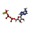


| #9: Chemical | ChemComp-AGS /  Mass: 523.247 Da / Num. of mol.: 4 / Source method: obtained synthetically / Formula: C10H16N5O12P3S / Feature type: SUBJECT OF INVESTIGATION / Comment: ATP-gamma-S, energy-carrying molecule analogue*YM Mass: 523.247 Da / Num. of mol.: 4 / Source method: obtained synthetically / Formula: C10H16N5O12P3S / Feature type: SUBJECT OF INVESTIGATION / Comment: ATP-gamma-S, energy-carrying molecule analogue*YM#10: Chemical | ChemComp-MG /  Mass: 24.305 Da / Num. of mol.: 4 / Source method: obtained synthetically / Formula: Mg / Feature type: SUBJECT OF INVESTIGATION Mass: 24.305 Da / Num. of mol.: 4 / Source method: obtained synthetically / Formula: Mg / Feature type: SUBJECT OF INVESTIGATION#11: Chemical | ChemComp-ADP / | Adenosine diphosphate Mass: 427.201 Da / Num. of mol.: 1 / Source method: obtained synthetically / Formula: C10H15N5O10P2 / Feature type: SUBJECT OF INVESTIGATION / Comment: ADP, energy-carrying molecule*YM Mass: 427.201 Da / Num. of mol.: 1 / Source method: obtained synthetically / Formula: C10H15N5O10P2 / Feature type: SUBJECT OF INVESTIGATION / Comment: ADP, energy-carrying molecule*YM |
|---|
-Details
| Has ligand of interest | Y |
|---|
-Experimental details
-Experiment
| Experiment | Method: ELECTRON MICROSCOPY |
|---|---|
| EM experiment | Aggregation state: PARTICLE / 3D reconstruction method: single particle reconstruction |
- Sample preparation
| Component | Name: (Replication Factor C RFC) bound to the sliding clamp (Proliferating Cell Nuclear Antigen PCNA) and primer-template DNA Type: COMPLEX / Entity ID: #1-#8 / Source: MULTIPLE SOURCES |
|---|---|
| Molecular weight | Value: 0.351 MDa / Experimental value: NO |
| Source (natural) | Organism: Saccharomyces cerevisiae (brewer's yeast) |
| Source (recombinant) | Organism: Escherichia coli BL21(DE3) (bacteria) |
| Buffer solution | pH: 7.5 |
| Specimen | Embedding applied: NO / Shadowing applied: NO / Staining applied: NO / Vitrification applied: YES |
| Vitrification | Cryogen name: ETHANE |
- Electron microscopy imaging
| Experimental equipment |  Model: Titan Krios / Image courtesy: FEI Company |
|---|---|
| Microscopy | Model: TFS KRIOS |
| Electron gun | Electron source: FIELD EMISSION GUN / Accelerating voltage: 300 kV / Illumination mode: FLOOD BEAM |
| Electron lens | Mode: BRIGHT FIELDBright-field microscopy / Calibrated magnification: 81000 X / Nominal defocus max: 2300 nm / Nominal defocus min: 1200 nm |
| Image recording | Electron dose: 40 e/Å2 / Film or detector model: GATAN K3 (6k x 4k) |
- Processing
| Software | Name: PHENIX / Version: dev_3699: / Classification: refinement | ||||||||||||||||||||||||
|---|---|---|---|---|---|---|---|---|---|---|---|---|---|---|---|---|---|---|---|---|---|---|---|---|---|
| EM software |
| ||||||||||||||||||||||||
| CTF correction | Type: PHASE FLIPPING AND AMPLITUDE CORRECTION | ||||||||||||||||||||||||
| Symmetry | Point symmetry: C1 (asymmetric) | ||||||||||||||||||||||||
| 3D reconstruction | Resolution: 3.4 Å / Resolution method: FSC 0.143 CUT-OFF / Num. of particles: 46300 / Symmetry type: POINT | ||||||||||||||||||||||||
| Atomic model building | Protocol: FLEXIBLE FIT / Space: REAL | ||||||||||||||||||||||||
| Atomic model building | PDB-ID: 1SXJ Accession code: 1SXJ / Source name: PDB / Type: experimental model | ||||||||||||||||||||||||
| Refine LS restraints |
|