 Movie
Movie Controller

[English] 日本語
 Yorodumi
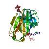
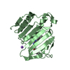
- PDB-7roj: Amyloid-related segment of alphaB-crystallin residues 90-100 with... -
+ Open data

- Basic information
| Entry | Database: PDB / ID: 7roj | ||||||
|---|---|---|---|---|---|---|---|
| Title | Amyloid-related segment of alphaB-crystallin residues 90-100 with G95W mutation | ||||||
 Components Components | Alpha-crystallin B chain peptide | ||||||
 Keywords Keywords | PROTEIN FIBRIL / amyloid oligomer | ||||||
| Function / homology |  Function and homology information Function and homology informationmicrotubule polymerization or depolymerization / negative regulation of intracellular transport / apoptotic process involved in morphogenesis / cardiac myofibril /  regulation of programmed cell death / tubulin complex assembly / structural constituent of eye lens / negative regulation of amyloid fibril formation / M band / lens development in camera-type eye ...microtubule polymerization or depolymerization / negative regulation of intracellular transport / apoptotic process involved in morphogenesis / cardiac myofibril / regulation of programmed cell death / tubulin complex assembly / structural constituent of eye lens / negative regulation of amyloid fibril formation / M band / lens development in camera-type eye / muscle organ development / actin filament bundle / HSF1-dependent transactivation / negative regulation of reactive oxygen species metabolic process / negative regulation of protein-containing complex assembly / stress-activated MAPK cascade / muscle contraction / synaptic membrane / response to hydrogen peroxide / cellular response to gamma radiation / negative regulation of cell growth / Z disc / unfolded protein binding / protein folding / response to estradiol / amyloid-beta binding / response to heat / perikaryon / protein refolding / microtubule binding / dendritic spine / lysosome / response to hypoxia / protein stabilization / axon / negative regulation of gene expression / negative regulation of DNA-templated transcription / protein-containing complex binding / negative regulation of apoptotic process / structural molecule activity / cell surface / protein homodimerization activity / protein-containing complex / mitochondrion / extracellular exosome / nucleoplasm / identical protein binding / metal ion binding / nucleus / cytosol / cytoplasm regulation of programmed cell death / tubulin complex assembly / structural constituent of eye lens / negative regulation of amyloid fibril formation / M band / lens development in camera-type eye ...microtubule polymerization or depolymerization / negative regulation of intracellular transport / apoptotic process involved in morphogenesis / cardiac myofibril / regulation of programmed cell death / tubulin complex assembly / structural constituent of eye lens / negative regulation of amyloid fibril formation / M band / lens development in camera-type eye / muscle organ development / actin filament bundle / HSF1-dependent transactivation / negative regulation of reactive oxygen species metabolic process / negative regulation of protein-containing complex assembly / stress-activated MAPK cascade / muscle contraction / synaptic membrane / response to hydrogen peroxide / cellular response to gamma radiation / negative regulation of cell growth / Z disc / unfolded protein binding / protein folding / response to estradiol / amyloid-beta binding / response to heat / perikaryon / protein refolding / microtubule binding / dendritic spine / lysosome / response to hypoxia / protein stabilization / axon / negative regulation of gene expression / negative regulation of DNA-templated transcription / protein-containing complex binding / negative regulation of apoptotic process / structural molecule activity / cell surface / protein homodimerization activity / protein-containing complex / mitochondrion / extracellular exosome / nucleoplasm / identical protein binding / metal ion binding / nucleus / cytosol / cytoplasmSimilarity search - Function | ||||||
| Biological species |  Homo sapiens (human) Homo sapiens (human) | ||||||
| Method | X-RAY DIFFRACTION / SYNCHROTRON / MOLECULAR REPLACEMENT / molecular replacement / Resolution: 1.6 Å | ||||||
| Model details | Amyloid Oligomer | ||||||
 Authors Authors | Sawaya, M.R. / Do, T.D. / Eisenberg, D.S. | ||||||
| Funding support |  United States, 1items United States, 1items
| ||||||
 Citation Citation | Journal: Protein Sci. / Year: 2022 Title: Atomic view of an amyloid dodecamer exhibiting selective cellular toxic vulnerability in acute brain slices. Authors: Gray, A.L.H. / Sawaya, M.R. / Acharyya, D. / Lou, J. / Edington, E.M. / Best, M.D. / Prosser, R.A. / Eisenberg, D.S. / Do, T.D. | ||||||
| History |
|
- Structure visualization
| Structure viewer | Molecule: MolmilJmol/JSmol |
|---|
- Downloads & links
-Download
| PDBx/mmCIF format | 7roj.cif.gz | 79.8 KB | Display | PDBx/mmCIF format |
|---|---|---|---|---|
| PDB format | pdb7roj.ent.gz | 53.2 KB | Display | PDB format |
| PDBx/mmJSON format | 7roj.json.gz | Tree view | PDBx/mmJSON format | |
| Others |  Other downloads Other downloads |
-Validation report
| Arichive directory | https://data.pdbj.org/pub/pdb/validation_reports/ro/7rojftp://data.pdbj.org/pub/pdb/validation_reports/ro/7roj | HTTPS FTP |
|---|
-Related structure data
| Related structure data |  7rolSC S: Starting model for refinement C: citing same article ( |
|---|---|
| Similar structure data |


-Links
 PDBj
PDBj

- Assembly
| Deposited unit | 
| ||||||||||||
|---|---|---|---|---|---|---|---|---|---|---|---|---|---|
| 1 |
| ||||||||||||
| Unit cell |
|
-Components
| #1: Protein/peptide | Mass: 1329.625 Da / Num. of mol.: 12 / Fragment: residues 90-100 / Mutation: G95W / Source method: obtained synthetically / Source: (synth.) Homo sapiens (human) / References: UniProt: P02511#2: Chemical | ChemComp-NA / |   Mass: 22.990 Da / Num. of mol.: 1 / Source method: obtained synthetically / Formula: Na Mass: 22.990 Da / Num. of mol.: 1 / Source method: obtained synthetically / Formula: Na#3: Chemical | ChemComp-TFA / Trifluoroacetic acid  Mass: 114.023 Da / Num. of mol.: 4 / Source method: obtained synthetically / Formula: C2HF3O2 Mass: 114.023 Da / Num. of mol.: 4 / Source method: obtained synthetically / Formula: C2HF3O2#4: Chemical | ChemComp-GOL / | Glycerol  Mass: 92.094 Da / Num. of mol.: 1 / Source method: obtained synthetically / Formula: C3H8O3 Mass: 92.094 Da / Num. of mol.: 1 / Source method: obtained synthetically / Formula: C3H8O3#5: Water | ChemComp-HOH / | Water Mass: 18.015 Da / Num. of mol.: 38 / Source method: isolated from a natural source / Formula: H2O Mass: 18.015 Da / Num. of mol.: 38 / Source method: isolated from a natural source / Formula: H2OHas ligand of interest | N | |
|---|
-Experimental details
-Experiment
| Experiment | Method: X-RAY DIFFRACTION / Number of used crystals: 1 |
|---|
- Sample preparation
| Crystal | Density Matthews: 1.84 Å3/Da / Density % sol: 33.3 % |
|---|---|
| Crystal grow | Temperature: 298 K / Method: vapor diffusion, hanging drop / pH: 7.5 Details: 1,4-butanediol, HEPES, NaCl, trifluoroacetic acid, glycerol |
-Data collection
| Diffraction | Mean temperature: 100 K / Serial crystal experiment: N | ||||||||||||||||||||||||||||||||||||||||||||||||||||||||||||||||||||||||||||||||||||||||||||||||||||||||||||||||||||||||||||||||||||||||||||||||||||||||||||||||||||||||
|---|---|---|---|---|---|---|---|---|---|---|---|---|---|---|---|---|---|---|---|---|---|---|---|---|---|---|---|---|---|---|---|---|---|---|---|---|---|---|---|---|---|---|---|---|---|---|---|---|---|---|---|---|---|---|---|---|---|---|---|---|---|---|---|---|---|---|---|---|---|---|---|---|---|---|---|---|---|---|---|---|---|---|---|---|---|---|---|---|---|---|---|---|---|---|---|---|---|---|---|---|---|---|---|---|---|---|---|---|---|---|---|---|---|---|---|---|---|---|---|---|---|---|---|---|---|---|---|---|---|---|---|---|---|---|---|---|---|---|---|---|---|---|---|---|---|---|---|---|---|---|---|---|---|---|---|---|---|---|---|---|---|---|---|---|---|---|---|---|---|
| Diffraction source | Source: SYNCHROTRON / Site: APS / Beamline: 24-ID-C / Wavelength: 0.97918 Å | ||||||||||||||||||||||||||||||||||||||||||||||||||||||||||||||||||||||||||||||||||||||||||||||||||||||||||||||||||||||||||||||||||||||||||||||||||||||||||||||||||||||||
| Detector | Type: DECTRIS EIGER2 X 16M / Detector: PIXEL / Date: Jul 31, 2020 | ||||||||||||||||||||||||||||||||||||||||||||||||||||||||||||||||||||||||||||||||||||||||||||||||||||||||||||||||||||||||||||||||||||||||||||||||||||||||||||||||||||||||
| Radiation | Monochromator: Si (111) / Protocol: SINGLE WAVELENGTH / Monochromatic (M) / Laue (L): M / Scattering type: x-ray | ||||||||||||||||||||||||||||||||||||||||||||||||||||||||||||||||||||||||||||||||||||||||||||||||||||||||||||||||||||||||||||||||||||||||||||||||||||||||||||||||||||||||
| Radiation wavelength | Wavelength: 0.97918 Å / Relative weight: 1 | ||||||||||||||||||||||||||||||||||||||||||||||||||||||||||||||||||||||||||||||||||||||||||||||||||||||||||||||||||||||||||||||||||||||||||||||||||||||||||||||||||||||||
| Reflection | Resolution: 1.6→64.09 Å / Num. obs: 14376 / % possible obs: 92.5 % / Redundancy: 2.042 % / Biso Wilson estimate: 32.292 Å2 / CC1/2: 0.995 / Rmerge(I) obs: 0.084 / Rrim(I) all: 0.111 / Χ2: 0.89 / Net I/σ(I): 6.14 | ||||||||||||||||||||||||||||||||||||||||||||||||||||||||||||||||||||||||||||||||||||||||||||||||||||||||||||||||||||||||||||||||||||||||||||||||||||||||||||||||||||||||
| Reflection shell | Diffraction-ID: 1
|
-Phasing
| Phasing | Method: molecular replacement | |||||||||
|---|---|---|---|---|---|---|---|---|---|---|
| Phasing MR | Model details: Phaser MODE: MR_AUTO
|
- Processing
| Software |
| |||||||||||||||||||||||||||||||||||||||||||||||||||||||||||||||||||||||||||||
|---|---|---|---|---|---|---|---|---|---|---|---|---|---|---|---|---|---|---|---|---|---|---|---|---|---|---|---|---|---|---|---|---|---|---|---|---|---|---|---|---|---|---|---|---|---|---|---|---|---|---|---|---|---|---|---|---|---|---|---|---|---|---|---|---|---|---|---|---|---|---|---|---|---|---|---|---|---|---|
| Refinement | Method to determine structure: MOLECULAR REPLACEMENT Starting model: 7ROL Resolution: 1.6→64.09 Å / SU ML: 0.2624 / Cross valid method: FREE R-VALUE / σ(F): 1.35 / Phase error: 34.9721 Stereochemistry target values: GeoStd + Monomer Library + CDL v1.2
| |||||||||||||||||||||||||||||||||||||||||||||||||||||||||||||||||||||||||||||
| Solvent computation | Shrinkage radii: 0.9 Å / VDW probe radii: 1.11 Å / Solvent model: FLAT BULK SOLVENT MODEL | |||||||||||||||||||||||||||||||||||||||||||||||||||||||||||||||||||||||||||||
| Displacement parameters | Biso mean: 33.78 Å2 | |||||||||||||||||||||||||||||||||||||||||||||||||||||||||||||||||||||||||||||
| Refinement step | Cycle: LAST / Resolution: 1.6→64.09 Å
| |||||||||||||||||||||||||||||||||||||||||||||||||||||||||||||||||||||||||||||
| Refine LS restraints |
| |||||||||||||||||||||||||||||||||||||||||||||||||||||||||||||||||||||||||||||
| LS refinement shell |
| |||||||||||||||||||||||||||||||||||||||||||||||||||||||||||||||||||||||||||||
| Refinement TLS params. | Method: refined / Origin x: 4.03843850142 Å / Origin y: -1.60843806554 Å / Origin z: 16.1599792453 Å
| |||||||||||||||||||||||||||||||||||||||||||||||||||||||||||||||||||||||||||||
| Refinement TLS group | Selection details: all |
