 Movie
Movie Controller


+ Open data

- Basic information
| Entry | Database: PDB / ID: 7rmp | ||||||
|---|---|---|---|---|---|---|---|
| Title | Structure of ACLY D1026A - substrates-asym | ||||||
 Components Components | ATP-citrate synthase | ||||||
 Keywords Keywords |  TRANSFERASE / mutant TRANSFERASE / mutant | ||||||
| Function / homology |  Function and homology informationATP citrate synthase activity / ATP citrate synthase / Fatty acyl-CoA biosynthesis / citrate metabolic process / ChREBP activates metabolic gene expression / acetyl-CoA biosynthetic process / oxaloacetate metabolic process / coenzyme A metabolic process / lipid biosynthetic process / cholesterol biosynthetic process ...ATP citrate synthase activity / ATP citrate synthase / Fatty acyl-CoA biosynthesis / citrate metabolic process / ChREBP activates metabolic gene expression / acetyl-CoA biosynthetic process / oxaloacetate metabolic process / coenzyme A metabolic process / lipid biosynthetic process / cholesterol biosynthetic process / tricarboxylic acid cycle / fatty acid biosynthetic process / azurophil granule lumen / ficolin-1-rich granule lumen / Neutrophil degranulation / extracellular exosome / extracellular region / nucleoplasm / ATP binding / membrane / metal ion binding / cytosol Function and homology informationATP citrate synthase activity / ATP citrate synthase / Fatty acyl-CoA biosynthesis / citrate metabolic process / ChREBP activates metabolic gene expression / acetyl-CoA biosynthetic process / oxaloacetate metabolic process / coenzyme A metabolic process / lipid biosynthetic process / cholesterol biosynthetic process ...ATP citrate synthase activity / ATP citrate synthase / Fatty acyl-CoA biosynthesis / citrate metabolic process / ChREBP activates metabolic gene expression / acetyl-CoA biosynthetic process / oxaloacetate metabolic process / coenzyme A metabolic process / lipid biosynthetic process / cholesterol biosynthetic process / tricarboxylic acid cycle / fatty acid biosynthetic process / azurophil granule lumen / ficolin-1-rich granule lumen / Neutrophil degranulation / extracellular exosome / extracellular region / nucleoplasm / ATP binding / membrane / metal ion binding / cytosolSimilarity search - Function | ||||||
| Biological species |  Homo sapiens (human) Homo sapiens (human) | ||||||
| Method | ELECTRON MICROSCOPY / single particle reconstruction / cryo EM / Resolution: 2.7 Å | ||||||
 Authors Authors | Wei, X. / Marmorstein, R. | ||||||
| Funding support |  United States, 1items United States, 1items
| ||||||
 Citation Citation | Journal: Nat Commun / Year: 2023 Title: Allosteric role of the citrate synthase homology domain of ATP citrate lyase. Authors: Xuepeng Wei / Kollin Schultz / Hannah L Pepper / Emily Megill / Austin Vogt / Nathaniel W Snyder / Ronen Marmorstein /  Abstract: ATP citrate lyase (ACLY) is the predominant nucleocytosolic source of acetyl-CoA and is aberrantly regulated in many diseases making it an attractive therapeutic target. Structural studies of ACLY ...ATP citrate lyase (ACLY) is the predominant nucleocytosolic source of acetyl-CoA and is aberrantly regulated in many diseases making it an attractive therapeutic target. Structural studies of ACLY reveal a central homotetrameric core citrate synthase homology (CSH) module flanked by acyl-CoA synthetase homology (ASH) domains, with ATP and citrate binding the ASH domain and CoA binding the ASH-CSH interface to produce acetyl-CoA and oxaloacetate products. The specific catalytic role of the CSH module and an essential D1026A residue contained within it has been a matter of debate. Here, we report biochemical and structural analysis of an ACLY-D1026A mutant demonstrating that this mutant traps a (3S)-citryl-CoA intermediate in the ASH domain in a configuration that is incompatible with the formation of acetyl-CoA, is able to convert acetyl-CoA and OAA to (3S)-citryl-CoA in the ASH domain, and can load CoA and unload acetyl-CoA in the CSH module. Together, this data support an allosteric role for the CSH module in ACLY catalysis. | ||||||
| History |
|
- Structure visualization
| Structure viewer | Molecule: MolmilJmol/JSmol |
|---|
- Downloads & links
-Download
| PDBx/mmCIF format | 7rmp.cif.gz | 713.1 KB | Display | PDBx/mmCIF format |
|---|---|---|---|---|
| PDB format | pdb7rmp.ent.gz | 583.7 KB | Display | PDB format |
| PDBx/mmJSON format | 7rmp.json.gz | Tree view | PDBx/mmJSON format | |
| Others |  Other downloads Other downloads |
-Validation report
| Arichive directory | https://data.pdbj.org/pub/pdb/validation_reports/rm/7rmpftp://data.pdbj.org/pub/pdb/validation_reports/rm/7rmp | HTTPS FTP |
|---|
-Related structure data
| Related structure data |  24577MC 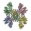 7rigC  7rkzC  8g1eC  8g1fC  8g5cC  8g5dC M: map data used to model this data C: citing same article ( |
|---|---|
| Similar structure data |


-Links
 PDBj
PDBj


- Assembly
| Deposited unit | 
|
|---|---|
| 1 |
|
-Components
-Protein , 1 types, 4 molecules ABCD
| #1: Protein | Mass: 120940.125 Da / Num. of mol.: 4 Source method: isolated from a genetically manipulated source Source: (gene. exp.) Homo sapiens (human) / Gene: ACLY / Production host:  Escherichia coli (E. coli) / References: UniProt: P53396, ATP citrate synthase Escherichia coli (E. coli) / References: UniProt: P53396, ATP citrate synthase |
|---|
-Non-polymers , 7 types, 60 molecules 


| #2: Chemical | Adenosine diphosphate Mass: 427.201 Da / Num. of mol.: 3 / Source method: obtained synthetically / Formula: C10H15N5O10P2 / Comment: ADP, energy-carrying molecule*YM Mass: 427.201 Da / Num. of mol.: 3 / Source method: obtained synthetically / Formula: C10H15N5O10P2 / Comment: ADP, energy-carrying molecule*YM#3: Chemical |  Mass: 941.642 Da / Num. of mol.: 3 / Source method: isolated from a natural source / Formula: C27H42N7O22P3S / Feature type: SUBJECT OF INVESTIGATION Mass: 941.642 Da / Num. of mol.: 3 / Source method: isolated from a natural source / Formula: C27H42N7O22P3S / Feature type: SUBJECT OF INVESTIGATION#4: Chemical | ChemComp-FLC / Citric acid Mass: 189.100 Da / Num. of mol.: 4 / Source method: obtained synthetically / Formula: C6H5O7 Mass: 189.100 Da / Num. of mol.: 4 / Source method: obtained synthetically / Formula: C6H5O7#5: Chemical | Mass: 767.534 Da / Num. of mol.: 2 / Source method: obtained synthetically #6: Chemical | ChemComp-COA / Coenzyme A Mass: 767.534 Da / Num. of mol.: 4 / Source method: obtained synthetically / Formula: C21H36N7O16P3S Mass: 767.534 Da / Num. of mol.: 4 / Source method: obtained synthetically / Formula: C21H36N7O16P3S#7: Chemical | Phosphate Mass: 94.971 Da / Num. of mol.: 3 / Source method: obtained synthetically / Formula: PO4 Mass: 94.971 Da / Num. of mol.: 3 / Source method: obtained synthetically / Formula: PO4#8: Water | ChemComp-HOH / | WaterMass: 18.015 Da / Num. of mol.: 41 / Source method: isolated from a natural source / Formula: H2O |
|---|
-Details
| Has ligand of interest | Y |
|---|
-Experimental details
-Experiment
| Experiment | Method: ELECTRON MICROSCOPY |
|---|---|
| EM experiment | Aggregation state: PARTICLE / 3D reconstruction method: single particle reconstruction |
- Sample preparation
| Component | Name: ACLY D1026A mutant in complex with CoA and citrate / Type: COMPLEX / Entity ID: #1 / Source: RECOMBINANT |
|---|---|
| Molecular weight | Value: 0.48 MDa / Experimental value: NO |
| Source (natural) | Organism: Homo sapiens (human) |
| Source (recombinant) | Organism: Escherichia coli (E. coli) |
| Buffer solution | pH: 7.5 |
| Specimen | Conc.: 4.5 mg/ml / Embedding applied: NO / Shadowing applied: NO / Staining applied: NO / Vitrification applied: YES |
| Vitrification | Cryogen name: ETHANE |
- Electron microscopy imaging
| Experimental equipment |  Model: Titan Krios / Image courtesy: FEI Company |
|---|---|
| Microscopy | Model: FEI TITAN KRIOS |
| Electron gun | Electron source: FIELD EMISSION GUN / Accelerating voltage: 300 kV / Illumination mode: FLOOD BEAM |
| Electron lens | Mode: BRIGHT FIELDBright-field microscopy |
| Image recording | Electron dose: 40 e/Å2 / Film or detector model: GATAN K3 (6k x 4k) |
- Processing
| Software | Name: PHENIX / Version: 1.19.2_4158: / Classification: refinement | ||||||||||||||||||||||||
|---|---|---|---|---|---|---|---|---|---|---|---|---|---|---|---|---|---|---|---|---|---|---|---|---|---|
| CTF correction | Type: PHASE FLIPPING AND AMPLITUDE CORRECTION | ||||||||||||||||||||||||
| 3D reconstruction | Resolution: 2.7 Å / Resolution method: FSC 0.143 CUT-OFF / Num. of particles: 100062 / Symmetry type: POINT | ||||||||||||||||||||||||
| Refine LS restraints |
|
