 Movie
Movie Controller

[English] 日本語
 Yorodumi
- PDB-7rar: Structure of Q67H mutant of disulfide stabilized HIV-1 CA hexamer -
+ Open data

- Basic information
| Entry | Database: PDB / ID: 7rar | ||||||||||||
|---|---|---|---|---|---|---|---|---|---|---|---|---|---|
| Title | Structure of Q67H mutant of disulfide stabilized HIV-1 CA hexamer | ||||||||||||
 Components Components | Capsid protein p24 | ||||||||||||
 Keywords Keywords | VIRAL PROTEIN | ||||||||||||
| Function / homology |  Function and homology information Function and homology informationviral budding via host ESCRT complex / ISG15 antiviral mechanism / host multivesicular body / viral nucleocapsid / host cell nucleus / host cell plasma membrane / virion membrane / structural molecule activity / RNA binding / zinc ion binding / membraneSimilarity search - Function | ||||||||||||
| Biological species |   Human immunodeficiency virus 1 Human immunodeficiency virus 1 | ||||||||||||
| Method | X-RAY DIFFRACTION / SYNCHROTRON / MOLECULAR REPLACEMENT / Resolution: 2.15 Å | ||||||||||||
 Authors Authors | Bester, S.M. / Kvaratskhelia, M. | ||||||||||||
| Funding support |  United States, 3items United States, 3items
| ||||||||||||
 Citation Citation | Journal: Mbio / Year: 2022 Title: Structural and Mechanistic Bases of Viral Resistance to HIV-1 Capsid Inhibitor Lenacapavir. Authors: Bester, S.M. / Adu-Ampratwum, D. / Annamalai, A.S. / Wei, G. / Briganti, L. / Murphy, B.C. / Haney, R. / Fuchs, J.R. / Kvaratskhelia, M. | ||||||||||||
| History |
|
- Structure visualization
| Structure viewer | Molecule: MolmilJmol/JSmol |
|---|
- Downloads & links
-Download
| PDBx/mmCIF format | 7rar.cif.gz | 120.7 KB | Display | PDBx/mmCIF format |
|---|---|---|---|---|
| PDB format | pdb7rar.ent.gz | 76.5 KB | Display | PDB format |
| PDBx/mmJSON format | 7rar.json.gz | Tree view | PDBx/mmJSON format | |
| Others |  Other downloads Other downloads |
-Validation report
| Arichive directory | https://data.pdbj.org/pub/pdb/validation_reports/ra/7rarftp://data.pdbj.org/pub/pdb/validation_reports/ra/7rar | HTTPS FTP |
|---|
-Related structure data
| Related structure data |  7rhmC  7rhnC  7rj2C  7rj4C  7rmjC  7rmmC 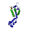 3h47S S: Starting model for refinement C: citing same article ( |
|---|---|
| Similar structure data |

-Links
 PDBj
PDBj

- Assembly
| Deposited unit | 
| ||||||||||||
|---|---|---|---|---|---|---|---|---|---|---|---|---|---|
| 1 | x 6
| ||||||||||||
| Unit cell |
|
-Components
| #1: Protein | / CA Mass: 25471.291 Da / Num. of mol.: 1 / Mutation: A14C, E45C, Q67H, W184A, M185A Source method: isolated from a genetically manipulated source Source: (gene. exp.) Human immunodeficiency virus 1 / Production host:  Escherichia coli BL21(DE3) (bacteria) / References: UniProt: P12493 Escherichia coli BL21(DE3) (bacteria) / References: UniProt: P12493 | ||||||
|---|---|---|---|---|---|---|---|
| #2: Chemical | ChemComp-IOD / Iodide  Mass: 126.904 Da / Num. of mol.: 6 / Source method: obtained synthetically / Formula: I Mass: 126.904 Da / Num. of mol.: 6 / Source method: obtained synthetically / Formula: I#3: Chemical | ChemComp-CL / Chloride  Mass: 35.453 Da / Num. of mol.: 4 / Source method: obtained synthetically / Formula: Cl Mass: 35.453 Da / Num. of mol.: 4 / Source method: obtained synthetically / Formula: Cl#4: Water | ChemComp-HOH / | Water Mass: 18.015 Da / Num. of mol.: 176 / Source method: isolated from a natural source / Formula: H2O Mass: 18.015 Da / Num. of mol.: 176 / Source method: isolated from a natural source / Formula: H2OHas ligand of interest | N | |
-Experimental details
-Experiment
| Experiment | Method: X-RAY DIFFRACTION / Number of used crystals: 1 |
|---|
- Sample preparation
| Crystal | Density Matthews: 2.76 Å3/Da / Density % sol: 55.47 % |
|---|---|
| Crystal grow | Temperature: 277.15 K / Method: vapor diffusion, hanging drop Details: 0.425M NaI, 4% peg 3350, 6% glycerol, 0.1M sodium cacodylate trihydrate pH 6.5 |
-Data collection
| Diffraction | Mean temperature: 100 K / Serial crystal experiment: N |
|---|---|
| Diffraction source | Source: SYNCHROTRON / Site: ALS / Beamline: 4.2.2 / Wavelength: 1.00003 Å |
| Detector | Type: RDI CMOS_8M / Detector: CMOS / Date: Oct 13, 2020 |
| Radiation | Protocol: SINGLE WAVELENGTH / Monochromatic (M) / Laue (L): M / Scattering type: x-ray |
| Radiation wavelength | Wavelength: 1.00003 Å / Relative weight: 1 |
| Reflection | Resolution: 2.15→46.75 Å / Num. obs: 15259 / % possible obs: 100 % / Redundancy: 10.8 % / Biso Wilson estimate: 27.73 Å2 / CC1/2: 0.997 / Rmerge(I) obs: 0.182 / Net I/σ(I): 12.2 |
| Reflection shell | Resolution: 2.15→2.22 Å / Redundancy: 10.2 % / Rmerge(I) obs: 1.59 / Mean I/σ(I) obs: 1.5 / Num. unique obs: 1325 / CC1/2: 0.667 / % possible all: 100 |
- Processing
| Software |
| ||||||||||||||||||||||||||||||||||||||||||||||||||||||||||||||||||||||||||||||||||||||||||||||||||||
|---|---|---|---|---|---|---|---|---|---|---|---|---|---|---|---|---|---|---|---|---|---|---|---|---|---|---|---|---|---|---|---|---|---|---|---|---|---|---|---|---|---|---|---|---|---|---|---|---|---|---|---|---|---|---|---|---|---|---|---|---|---|---|---|---|---|---|---|---|---|---|---|---|---|---|---|---|---|---|---|---|---|---|---|---|---|---|---|---|---|---|---|---|---|---|---|---|---|---|---|---|---|
| Refinement | Method to determine structure: MOLECULAR REPLACEMENT Starting model: 3H47 Resolution: 2.15→46.75 Å / SU ML: 0.2285 / Cross valid method: FREE R-VALUE / σ(F): 1.34 / Phase error: 26.6613 Stereochemistry target values: GeoStd + Monomer Library + CDL v1.2
| ||||||||||||||||||||||||||||||||||||||||||||||||||||||||||||||||||||||||||||||||||||||||||||||||||||
| Solvent computation | Shrinkage radii: 0.9 Å / VDW probe radii: 1.11 Å / Solvent model: FLAT BULK SOLVENT MODEL | ||||||||||||||||||||||||||||||||||||||||||||||||||||||||||||||||||||||||||||||||||||||||||||||||||||
| Displacement parameters | Biso mean: 35.65 Å2 | ||||||||||||||||||||||||||||||||||||||||||||||||||||||||||||||||||||||||||||||||||||||||||||||||||||
| Refinement step | Cycle: LAST / Resolution: 2.15→46.75 Å
| ||||||||||||||||||||||||||||||||||||||||||||||||||||||||||||||||||||||||||||||||||||||||||||||||||||
| Refine LS restraints |
| ||||||||||||||||||||||||||||||||||||||||||||||||||||||||||||||||||||||||||||||||||||||||||||||||||||
| LS refinement shell |
| ||||||||||||||||||||||||||||||||||||||||||||||||||||||||||||||||||||||||||||||||||||||||||||||||||||
| Refinement TLS params. | Method: refined / Refine-ID: X-RAY DIFFRACTION
| ||||||||||||||||||||||||||||||||||||||||||||||||||||||||||||||||||||||||||||||||||||||||||||||||||||
| Refinement TLS group | Refine-ID: X-RAY DIFFRACTION / Auth asym-ID: C / Label asym-ID: A
|
