 Movie
Movie Controller


+ Open data

- Basic information
| Entry | Database: PDB / ID: 7r94 | |||||||||
|---|---|---|---|---|---|---|---|---|---|---|
| Title | T-Plastin-F-actin complex | |||||||||
 Components Components |
| |||||||||
 Keywords Keywords |  STRUCTURAL PROTEIN / T-plastin / plastin / actin / filopodium / cytoskeleton / cell protrusion STRUCTURAL PROTEIN / T-plastin / plastin / actin / filopodium / cytoskeleton / cell protrusion | |||||||||
| Function / homology |  Function and homology information Function and homology informationactin filament network formation / Striated Muscle Contraction / actin filament bundle / actin filament bundle assembly / skeletal muscle thin filament assembly / striated muscle thin filament / skeletal muscle fiber development / stress fiber / actin filament / Hydrolases; Acting on acid anhydrides; Acting on acid anhydrides to facilitate cellular and subcellular movement ...actin filament network formation / Striated Muscle Contraction / actin filament bundle / actin filament bundle assembly / skeletal muscle thin filament assembly / striated muscle thin filament / skeletal muscle fiber development / stress fiber / actin filament / Hydrolases; Acting on acid anhydrides; Acting on acid anhydrides to facilitate cellular and subcellular movement / bone development / actin filament binding / hydrolase activity / calcium ion binding / ATP binding / plasma membrane / cytosol / cytoplasmSimilarity search - Function | |||||||||
| Biological species |  Homo sapiens (human) Homo sapiens (human) Gallus gallus (chicken) Gallus gallus (chicken) | |||||||||
| Method | ELECTRON MICROSCOPY / helical reconstruction / cryo EM / Resolution: 2.6 Å | |||||||||
 Authors Authors | Mei, L. / Alushin, G.M. | |||||||||
| Funding support |  United States, 2items United States, 2items
| |||||||||
 Citation Citation | Journal: Proc Natl Acad Sci U S A / Year: 2022 Title: Structural mechanism for bidirectional actin cross-linking by T-plastin. Authors: Lin Mei / Matthew J Reynolds / Damien Garbett / Rui Gong / Tobias Meyer / Gregory M Alushin / Abstract: To orchestrate cell mechanics, trafficking, and motility, cytoskeletal filaments must assemble into higher-order networks whose local subcellular architecture and composition specify their functions. ...To orchestrate cell mechanics, trafficking, and motility, cytoskeletal filaments must assemble into higher-order networks whose local subcellular architecture and composition specify their functions. Cross-linking proteins bridge filaments at the nanoscale to control a network's μm-scale geometry, thereby conferring its mechanical properties and functional dynamics. While these interfilament linkages are key determinants of cytoskeletal function, their structural mechanisms remain poorly understood. Plastins/fimbrins are an evolutionarily ancient family of tandem calponin-homology domain (CHD) proteins required to construct multiple classes of actin networks, which feature diverse geometries specialized to power cytokinesis, microvilli and stereocilia biogenesis, and persistent cell migration. Here, we focus on the structural basis of actin network assembly by human T-plastin, a ubiquitously expressed isoform necessary for the maintenance of stable cellular protrusions generated by actin polymerization forces. By implementing a machine-learning-enabled cryo-electron microscopy pipeline for visualizing cross-linkers bridging multiple filaments, we uncover a sequential bundling mechanism enabling T-plastin to bridge pairs of actin filaments in both parallel and antiparallel orientations. T-plastin populates distinct structural landscapes in these two bridging orientations that are selectively compatible with actin networks featuring divergent architectures and functions. Our structural, biochemical, and cell biological data highlight inter-CHD linkers as key structural elements underlying flexible but stable cross-linking that are likely to be disrupted by T-plastin mutations that cause hereditary bone diseases. | |||||||||
| History |
|
- Structure visualization
| Structure viewer | Molecule: MolmilJmol/JSmol |
|---|
- Downloads & links
-Download
| PDBx/mmCIF format | 7r94.cif.gz | 449.7 KB | Display | PDBx/mmCIF format |
|---|---|---|---|---|
| PDB format | pdb7r94.ent.gz | 369.1 KB | Display | PDB format |
| PDBx/mmJSON format | 7r94.json.gz | Tree view | PDBx/mmJSON format | |
| Others |  Other downloads Other downloads |
-Validation report
| Arichive directory | https://data.pdbj.org/pub/pdb/validation_reports/r9/7r94ftp://data.pdbj.org/pub/pdb/validation_reports/r9/7r94 | HTTPS FTP |
|---|
-Related structure data
| Related structure data |  24323MC 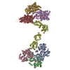 7sx8C  7sx9C  7sxaC M: map data used to model this data C: citing same article ( |
|---|---|
| Similar structure data |


-Links
 PDBj
PDBj


- Assembly
| Deposited unit | 
|
|---|---|
| 1 |
|
-Components
| #1: Protein | / Alpha-actin-1 Mass: 42109.973 Da / Num. of mol.: 5 / Source method: isolated from a natural source / Source: (natural) Gallus gallus (chicken) / References: UniProt: P68139#2: Protein | / T-plastinMass: 70896.867 Da / Num. of mol.: 3 Source method: isolated from a genetically manipulated source Source: (gene. exp.) Homo sapiens (human) / Gene: PLS3 / Production host:  Escherichia coli (E. coli) / References: UniProt: P13797 Escherichia coli (E. coli) / References: UniProt: P13797#3: Chemical | ChemComp-ADP / Adenosine diphosphate  Mass: 427.201 Da / Num. of mol.: 5 / Source method: obtained synthetically / Formula: C10H15N5O10P2 / Comment: ADP, energy-carrying molecule*YM Mass: 427.201 Da / Num. of mol.: 5 / Source method: obtained synthetically / Formula: C10H15N5O10P2 / Comment: ADP, energy-carrying molecule*YM#4: Chemical | ChemComp-MG /   Mass: 24.305 Da / Num. of mol.: 5 / Source method: obtained synthetically / Formula: Mg Mass: 24.305 Da / Num. of mol.: 5 / Source method: obtained synthetically / Formula: MgHas ligand of interest | N | |
|---|
-Experimental details
-Experiment
| Experiment | Method: ELECTRON MICROSCOPY |
|---|---|
| EM experiment | Aggregation state: HELICAL ARRAY / 3D reconstruction method: helical reconstruction |
- Sample preparation
| Component | Name: T-Plastin-F-actin complex / Type: COMPLEX Details: Full-length human T-Plastin (PLS3) bound to F-actin in the presence of calcium Entity ID: #1-#2 / Source: MULTIPLE SOURCES |
|---|---|
| Molecular weight | Value: 27.5 kDa/nm / Experimental value: NO |
| Source (natural) | Organism: Homo sapiens (human) |
| Buffer solution | pH: 7.5 |
| Specimen | Embedding applied: NO / Shadowing applied: NO / Staining applied: NO / Vitrification applied: YES |
| Vitrification | Cryogen name: ETHANE |
- Electron microscopy imaging
| Experimental equipment |  Model: Titan Krios / Image courtesy: FEI Company |
|---|---|
| Microscopy | Model: FEI TITAN KRIOS |
| Electron gun | Electron source: FIELD EMISSION GUN / Accelerating voltage: 300 kV / Illumination mode: FLOOD BEAM |
| Electron lens | Mode: BRIGHT FIELDBright-field microscopy |
| Image recording | Average exposure time: 10 sec. / Electron dose: 71.02 e/Å2 / Detector mode: COUNTING / Film or detector model: GATAN K2 SUMMIT (4k x 4k) / Num. of grids imaged: 1 |
| Image scans | Movie frames/image: 50 / Used frames/image: 1-50 |
- Processing
| EM software |
| ||||||||||||||||||||||||||||||||||||||||
|---|---|---|---|---|---|---|---|---|---|---|---|---|---|---|---|---|---|---|---|---|---|---|---|---|---|---|---|---|---|---|---|---|---|---|---|---|---|---|---|---|---|
| CTF correction | Type: PHASE FLIPPING AND AMPLITUDE CORRECTION | ||||||||||||||||||||||||||||||||||||||||
| Helical symmerty | Angular rotation/subunit: -166.8 ° / Axial rise/subunit: 27.4 Å / Axial symmetry: C1 | ||||||||||||||||||||||||||||||||||||||||
| Particle selection | Num. of particles selected: 577659 / Details: helical auto-picking | ||||||||||||||||||||||||||||||||||||||||
| 3D reconstruction | Resolution: 2.6 Å / Resolution method: FSC 0.143 CUT-OFF / Num. of particles: 442523 / Symmetry type: HELICAL | ||||||||||||||||||||||||||||||||||||||||
| Atomic model building | PDB-ID: 6UPV Pdb chain-ID: A |

