 Movie
Movie Controller

[English] 日本語
 Yorodumi
- PDB-7qrf: Structure of the dimeric complex between precursor membrane ectod... -
+ Open data

- Basic information
| Entry | Database: PDB / ID: 7qrf | ||||||||||||
|---|---|---|---|---|---|---|---|---|---|---|---|---|---|
| Title | Structure of the dimeric complex between precursor membrane ectodomain (prM) and envelope protein ectodomain (E) from tick-borne encephalitis virus | ||||||||||||
 Components Components |
| ||||||||||||
 Keywords Keywords |  VIRAL PROTEIN / class II fusion envelope protein / precursor membrane protein / chaperone / flavivirus maturation / trans-golgi acid pH VIRAL PROTEIN / class II fusion envelope protein / precursor membrane protein / chaperone / flavivirus maturation / trans-golgi acid pH | ||||||||||||
| Function / homology |  Function and homology informationflavivirin / symbiont-mediated suppression of host JAK-STAT cascade via inhibition of STAT2 activity / symbiont-mediated suppression of host JAK-STAT cascade via inhibition of STAT1 activity / viral capsid / double-stranded RNA binding / nucleoside-triphosphate phosphatase / mRNA (guanine-N7)-methyltransferase / methyltransferase cap1 / clathrin-dependent endocytosis of virus by host cell / mRNA (nucleoside-2'-O-)-methyltransferase activity ...flavivirin / symbiont-mediated suppression of host JAK-STAT cascade via inhibition of STAT2 activity / symbiont-mediated suppression of host JAK-STAT cascade via inhibition of STAT1 activity / viral capsid / double-stranded RNA binding / nucleoside-triphosphate phosphatase / mRNA (guanine-N7)-methyltransferase / methyltransferase cap1 / clathrin-dependent endocytosis of virus by host cell / mRNA (nucleoside-2'-O-)-methyltransferase activity / mRNA 5'-cap (guanine-N7-)-methyltransferase activity / RNA helicase activity / host cell endoplasmic reticulum membrane / host cell perinuclear region of cytoplasm / protein dimerization activity / RNA helicase / induction by virus of host autophagy / RNA-directed RNA polymerase / viral RNA genome replication / RNA-dependent RNA polymerase activity / serine-type endopeptidase activity / fusion of virus membrane with host endosome membrane / viral envelope / symbiont-mediated suppression of host type I interferon-mediated signaling pathway / host cell nucleus / virion attachment to host cell / virion membrane / structural molecule activity / ATP hydrolysis activity / proteolysis / extracellular region / ATP binding / membrane / metal ion binding Function and homology informationflavivirin / symbiont-mediated suppression of host JAK-STAT cascade via inhibition of STAT2 activity / symbiont-mediated suppression of host JAK-STAT cascade via inhibition of STAT1 activity / viral capsid / double-stranded RNA binding / nucleoside-triphosphate phosphatase / mRNA (guanine-N7)-methyltransferase / methyltransferase cap1 / clathrin-dependent endocytosis of virus by host cell / mRNA (nucleoside-2'-O-)-methyltransferase activity ...flavivirin / symbiont-mediated suppression of host JAK-STAT cascade via inhibition of STAT2 activity / symbiont-mediated suppression of host JAK-STAT cascade via inhibition of STAT1 activity / viral capsid / double-stranded RNA binding / nucleoside-triphosphate phosphatase / mRNA (guanine-N7)-methyltransferase / methyltransferase cap1 / clathrin-dependent endocytosis of virus by host cell / mRNA (nucleoside-2'-O-)-methyltransferase activity / mRNA 5'-cap (guanine-N7-)-methyltransferase activity / RNA helicase activity / host cell endoplasmic reticulum membrane / host cell perinuclear region of cytoplasm / protein dimerization activity / RNA helicase / induction by virus of host autophagy / RNA-directed RNA polymerase / viral RNA genome replication / RNA-dependent RNA polymerase activity / serine-type endopeptidase activity / fusion of virus membrane with host endosome membrane / viral envelope / symbiont-mediated suppression of host type I interferon-mediated signaling pathway / host cell nucleus / virion attachment to host cell / virion membrane / structural molecule activity / ATP hydrolysis activity / proteolysis / extracellular region / ATP binding / membrane / metal ion bindingSimilarity search - Function | ||||||||||||
| Biological species |  Tick-borne encephalitis virus Tick-borne encephalitis virus | ||||||||||||
| Method | X-RAY DIFFRACTION / SYNCHROTRON / MOLECULAR REPLACEMENT / Resolution: 2.28 Å | ||||||||||||
 Authors Authors | Vaney, M.C. / Rouvinski, A. / Rey, F.A. | ||||||||||||
| Funding support |  France, 3items France, 3items
| ||||||||||||
 Citation Citation | Journal: Nat Commun / Year: 2022 Title: Evolution and activation mechanism of the flavivirus class II membrane-fusion machinery. Authors: Vaney, M.C. / Dellarole, M. / Duquerroy, S. / Medits, I. / Tsouchnikas, G. / Rouvinski, A. / England, P. / Stiasny, K. / Heinz, F.X. / Rey, F.A. #1: Journal: Nature / Year: 1995Title: The envelope glycoprotein from tick-borne encephalitis virus at 2 A resolution. Authors: Rey, F.A. / Heinz, F.X. / Mandl, C. / Kunz, C. / Harrison, S.C. | ||||||||||||
| History |
|
- Structure visualization
| Structure viewer | Molecule: MolmilJmol/JSmol |
|---|
- Downloads & links
-Download
| PDBx/mmCIF format | 7qrf.cif.gz | 205.5 KB | Display | PDBx/mmCIF format |
|---|---|---|---|---|
| PDB format | pdb7qrf.ent.gz | 161.1 KB | Display | PDB format |
| PDBx/mmJSON format | 7qrf.json.gz | Tree view | PDBx/mmJSON format | |
| Others |  Other downloads Other downloads |
-Validation report
| Arichive directory | https://data.pdbj.org/pub/pdb/validation_reports/qr/7qrfftp://data.pdbj.org/pub/pdb/validation_reports/qr/7qrf | HTTPS FTP |
|---|
-Related structure data
| Related structure data |  7qreC 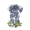 7qrgC 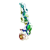 1svbS  3c5xS C: citing same article ( S: Starting model for refinement |
|---|---|
| Similar structure data |
-Links
 PDBj
PDBj


- Assembly
| Deposited unit | 
| ||||||||
|---|---|---|---|---|---|---|---|---|---|
| 1 | 
| ||||||||
| Unit cell |
| ||||||||
| Components on special symmetry positions |
|
-Components
-Protein , 2 types, 2 molecules AD
| #1: Protein | Mass: 47867.855 Da / Num. of mol.: 1 Source method: isolated from a genetically manipulated source Details: The N-terminal end of E (Molecule 1) is linked to the C-terminal of ectodomain prM (Molecule 3) by a linker of 14 residues (GGGGENLYFQGGGG). This linker of 14 residues is only described in ...Details: The N-terminal end of E (Molecule 1) is linked to the C-terminal of ectodomain prM (Molecule 3) by a linker of 14 residues (GGGGENLYFQGGGG). This linker of 14 residues is only described in the sequence of E (Molecule 1) at its N-terminal. For purification, at the C-terminal of E it is added an enterokinase site with a dstrep-tag sequence : (GPFEDDDDKAGWSHPQFEKGGGSGGGSGGGSWSHPQFEK). Source: (gene. exp.) Tick-borne encephalitis virus (WESTERN SUBTYPE)Strain: Neudoerfl / Plasmid: PT389 / Cell (production host): SCHNEIDER 2 / Production host:  Drosophila melanogaster (fruit fly) / References: UniProt: P14336 Drosophila melanogaster (fruit fly) / References: UniProt: P14336 |
|---|---|
| #3: Protein | Mass: 15290.941 Da / Num. of mol.: 1 Source method: isolated from a genetically manipulated source Details: The C-terminal end of prM (Molecule 3) is linked to the N-terminal of E (Molecule 1) by a linker of 14 residues (GGGGENLYFQGGGG). This linker is described in the sequence of E (Molecule 1). ...Details: The C-terminal end of prM (Molecule 3) is linked to the N-terminal of E (Molecule 1) by a linker of 14 residues (GGGGENLYFQGGGG). This linker is described in the sequence of E (Molecule 1). The furin site is deleted from one amino acid (Arginine). Intact furin site : ...RTRRSVL... Mutated furin site : ...RTRSVL... Source: (gene. exp.) Tick-borne encephalitis virus (WESTERN SUBTYPE)Strain: Neudoerfl / Plasmid: PT389 / Cell (production host): SCHNEIDER 2 / Production host: Drosophila melanogaster (fruit fly) / References: UniProt: P14336 |
-Protein/peptide , 1 types, 1 molecules C
| #2: Protein/peptide | Mass: 443.539 Da / Num. of mol.: 1 Source method: isolated from a genetically manipulated source Details: This peptide should be part of the M ectodomain but we were unable to attribute the true sequence. Source: (gene. exp.) Tick-borne encephalitis virus (WESTERN SUBTYPE)Strain: NEUDOERFL / Plasmid: PT389 / Cell (production host): SCHNEIDER 2 / Production host: Drosophila melanogaster (fruit fly) |
|---|
-Non-polymers , 4 types, 84 molecules 


| #4: Chemical | ChemComp-SO4 / Sulfate Mass: 96.063 Da / Num. of mol.: 1 / Source method: obtained synthetically / Formula: SO4 Mass: 96.063 Da / Num. of mol.: 1 / Source method: obtained synthetically / Formula: SO4 | ||
|---|---|---|---|
| #5: Chemical | ChemComp-PE4 / Polyethylene glycol Mass: 354.436 Da / Num. of mol.: 1 / Source method: obtained synthetically / Formula: C16H34O8 / Comment: precipitant*YM Mass: 354.436 Da / Num. of mol.: 1 / Source method: obtained synthetically / Formula: C16H34O8 / Comment: precipitant*YM | ||
| #6: Chemical | ChemComp-EDO / Ethylene glycol Mass: 62.068 Da / Num. of mol.: 8 / Source method: obtained synthetically / Formula: C2H6O2 Mass: 62.068 Da / Num. of mol.: 8 / Source method: obtained synthetically / Formula: C2H6O2#7: Water | ChemComp-HOH / | WaterMass: 18.015 Da / Num. of mol.: 74 / Source method: isolated from a natural source / Formula: H2O |
-Details
| Has ligand of interest | N |
|---|
-Experimental details
-Experiment
| Experiment | Method: X-RAY DIFFRACTION / Number of used crystals: 1 |
|---|
- Sample preparation
| Crystal | Density Matthews: 2.97 Å3/Da / Density % sol: 58.63 % |
|---|---|
| Crystal grow | Temperature: 293 K / Method: vapor diffusion / pH: 3.5 Details: 0.2M Li(SO4), 0.1M Na citrate pH 3.5, 28% (v/v) PEG 400 |
-Data collection
| Diffraction | Mean temperature: 100 K / Serial crystal experiment: N |
|---|---|
| Diffraction source | Source: SYNCHROTRON / Site: ESRF / Beamline: ID23-1 / Wavelength: 0.97934 Å |
| Detector | Type: DECTRIS PILATUS 6M / Detector: PIXEL / Date: Feb 14, 2014 |
| Radiation | Protocol: SINGLE WAVELENGTH / Monochromatic (M) / Laue (L): M / Scattering type: x-ray |
| Radiation wavelength | Wavelength: 0.97934 Å / Relative weight: 1 |
| Reflection | Resolution: 2.28→49 Å / Num. obs: 28985 / % possible obs: 95 % / Redundancy: 19.5 % / CC1/2: 0.998 / Rmerge(I) obs: 0.096 / Rpim(I) all: 0.023 / Rrim(I) all: 0.099 / Net I/σ(I): 19.8 |
| Reflection shell | Resolution: 2.28→2.435 Å / Rmerge(I) obs: 2.623 / Num. unique obs: 1449 / CC1/2: 0.323 / Rpim(I) all: 0.891 / Rrim(I) all: 2.699 |
- Processing
| Software |
| |||||||||||||||||||||||||||||||||||||||||||||||||||||||||||||||||||||||||||||||||||||||||||||||||||||||||||||||||||||||||||||||||||||||||||||||||||||||||||||||||||||||||||||||
|---|---|---|---|---|---|---|---|---|---|---|---|---|---|---|---|---|---|---|---|---|---|---|---|---|---|---|---|---|---|---|---|---|---|---|---|---|---|---|---|---|---|---|---|---|---|---|---|---|---|---|---|---|---|---|---|---|---|---|---|---|---|---|---|---|---|---|---|---|---|---|---|---|---|---|---|---|---|---|---|---|---|---|---|---|---|---|---|---|---|---|---|---|---|---|---|---|---|---|---|---|---|---|---|---|---|---|---|---|---|---|---|---|---|---|---|---|---|---|---|---|---|---|---|---|---|---|---|---|---|---|---|---|---|---|---|---|---|---|---|---|---|---|---|---|---|---|---|---|---|---|---|---|---|---|---|---|---|---|---|---|---|---|---|---|---|---|---|---|---|---|---|---|---|---|---|---|
| Refinement | Method to determine structure: MOLECULAR REPLACEMENT Starting model: 1SVB, 3C5X Resolution: 2.28→28.42 Å / Cor.coef. Fo:Fc: 0.96 / Cor.coef. Fo:Fc free: 0.939 / SU R Cruickshank DPI: 0.222 / Cross valid method: THROUGHOUT / σ(F): 0 / SU R Blow DPI: 0.217 / SU Rfree Blow DPI: 0.18 / SU Rfree Cruickshank DPI: 0.183 Details: The BUSTER refinement was done against the STARANISO corrected intensity from anisotropy. This is the reason why, in the last shell of resolution, the completness is 7% while the working ...Details: The BUSTER refinement was done against the STARANISO corrected intensity from anisotropy. This is the reason why, in the last shell of resolution, the completness is 7% while the working Rfactor is higher than the free Rfactor. All the refinement stastitics introduced here are from the STARANISO statistics output.
| |||||||||||||||||||||||||||||||||||||||||||||||||||||||||||||||||||||||||||||||||||||||||||||||||||||||||||||||||||||||||||||||||||||||||||||||||||||||||||||||||||||||||||||||
| Displacement parameters | Biso max: 178.25 Å2 / Biso mean: 80.83 Å2 / Biso min: 44.08 Å2
| |||||||||||||||||||||||||||||||||||||||||||||||||||||||||||||||||||||||||||||||||||||||||||||||||||||||||||||||||||||||||||||||||||||||||||||||||||||||||||||||||||||||||||||||
| Refine analyze | Luzzati coordinate error obs: 0.3 Å | |||||||||||||||||||||||||||||||||||||||||||||||||||||||||||||||||||||||||||||||||||||||||||||||||||||||||||||||||||||||||||||||||||||||||||||||||||||||||||||||||||||||||||||||
| Refinement step | Cycle: final / Resolution: 2.28→28.42 Å
| |||||||||||||||||||||||||||||||||||||||||||||||||||||||||||||||||||||||||||||||||||||||||||||||||||||||||||||||||||||||||||||||||||||||||||||||||||||||||||||||||||||||||||||||
| Refine LS restraints |
| |||||||||||||||||||||||||||||||||||||||||||||||||||||||||||||||||||||||||||||||||||||||||||||||||||||||||||||||||||||||||||||||||||||||||||||||||||||||||||||||||||||||||||||||
| LS refinement shell | Resolution: 2.28→2.36 Å / Rfactor Rfree error: 0 / Total num. of bins used: 15
| |||||||||||||||||||||||||||||||||||||||||||||||||||||||||||||||||||||||||||||||||||||||||||||||||||||||||||||||||||||||||||||||||||||||||||||||||||||||||||||||||||||||||||||||
| Refinement TLS params. | Method: refined / Refine-ID: X-RAY DIFFRACTION
| |||||||||||||||||||||||||||||||||||||||||||||||||||||||||||||||||||||||||||||||||||||||||||||||||||||||||||||||||||||||||||||||||||||||||||||||||||||||||||||||||||||||||||||||
| Refinement TLS group |
|
