 Movie
Movie Controller

[English] 日本語
 Yorodumi
- PDB-7qjn: Crystal structure of an alpha/beta-hydrolase enzyme from Candidat... -
+ Open data

- Basic information
| Entry | Database: PDB / ID: 7qjn | ||||||
|---|---|---|---|---|---|---|---|
| Title | Crystal structure of an alpha/beta-hydrolase enzyme from Candidatus Kryptobacter tengchongensis (306) | ||||||
 Components Components | Dienelactone hydrolase | ||||||
 Keywords Keywords |  HYDROLASE / plastic degradation HYDROLASE / plastic degradation | ||||||
| Function / homology | Dienelactone hydrolase / Dienelactone hydrolase family / Alpha/Beta hydrolase fold / hydrolase activity / PHOSPHATE ION / Dienelactone hydrolase Function and homology information Function and homology information | ||||||
| Biological species |  Candidatus Kryptobacter tengchongensis (bacteria) Candidatus Kryptobacter tengchongensis (bacteria) | ||||||
| Method | X-RAY DIFFRACTION / SYNCHROTRON / MOLECULAR REPLACEMENT / Resolution: 1.885 Å | ||||||
 Authors Authors | Zahn, M. / Gill, R.S. / Erickson, E. / Beckham, G.T. / McGeehan, J.E. | ||||||
| Funding support |  United Kingdom, 1items United Kingdom, 1items
| ||||||
 Citation Citation | Journal: Nat Commun / Year: 2022 Title: Sourcing thermotolerant poly(ethylene terephthalate) hydrolase scaffolds from natural diversity Authors: Erickson, E. / Gado, J.E. / Avilan, L. / Bratti, F. / Brizendine, R.K. / Cox, P.A. / Gill, R. / Graham, R. / Kim, D.J. / Konig, G. / Michener, W.E. / Poudel, S. / Ramirez, K.J. / ...Authors: Erickson, E. / Gado, J.E. / Avilan, L. / Bratti, F. / Brizendine, R.K. / Cox, P.A. / Gill, R. / Graham, R. / Kim, D.J. / Konig, G. / Michener, W.E. / Poudel, S. / Ramirez, K.J. / Shakespeare, T.J. / Zahn, M. / Boyd, E.S. / Payne, C.M. / DuBois, J.L. / Pickford, A.R. / Beckham, G.T. / McGeehan, J.E. | ||||||
| History |
|
- Structure visualization
| Structure viewer | Molecule: MolmilJmol/JSmol |
|---|
- Downloads & links
-Download
| PDBx/mmCIF format | 7qjn.cif.gz | 127.4 KB | Display | PDBx/mmCIF format |
|---|---|---|---|---|
| PDB format | pdb7qjn.ent.gz | 98.6 KB | Display | PDB format |
| PDBx/mmJSON format | 7qjn.json.gz | Tree view | PDBx/mmJSON format | |
| Others |  Other downloads Other downloads |
-Validation report
| Arichive directory | https://data.pdbj.org/pub/pdb/validation_reports/qj/7qjnftp://data.pdbj.org/pub/pdb/validation_reports/qj/7qjn | HTTPS FTP |
|---|
-Related structure data
| Related structure data |  7qjmC 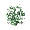 7qjoC  7qjpC  7qjqC  7qjrC  7qjsC  7qjtC C: citing same article ( |
|---|---|
| Similar structure data |
-Links
 PDBj
PDBj- Assembly
| Deposited unit | 
| ||||||||
|---|---|---|---|---|---|---|---|---|---|
| 1 |
| ||||||||
| Unit cell |
| ||||||||
| Components on special symmetry positions |
|
-Components
| #1: Protein | Mass: 33135.820 Da / Num. of mol.: 1 Source method: isolated from a genetically manipulated source Details: alpha/beta-hydrolase Source: (gene. exp.) Candidatus Kryptobacter tengchongensis (bacteria)Gene: JGI24_00892 / Production host: Escherichia coli BL21(DE3) (bacteria) / References: UniProt: A0A656D8B6 | ||||
|---|---|---|---|---|---|
| #2: Chemical | Phosphate  Mass: 94.971 Da / Num. of mol.: 3 / Source method: obtained synthetically / Formula: PO4 / Feature type: SUBJECT OF INVESTIGATION Mass: 94.971 Da / Num. of mol.: 3 / Source method: obtained synthetically / Formula: PO4 / Feature type: SUBJECT OF INVESTIGATION#3: Water | ChemComp-HOH / | Water Mass: 18.015 Da / Num. of mol.: 108 / Source method: isolated from a natural source / Formula: H2O Mass: 18.015 Da / Num. of mol.: 108 / Source method: isolated from a natural source / Formula: H2OHas ligand of interest | Y | |
-Experimental details
-Experiment
| Experiment | Method: X-RAY DIFFRACTION / Number of used crystals: 1 |
|---|
- Sample preparation
| Crystal | Density Matthews: 2.85 Å3/Da / Density % sol: 56.82 % |
|---|---|
| Crystal grow | Temperature: 293 K / Method: vapor diffusion, sitting drop Details: 1.8 M sodium phosphate monobasic monohydrate / potassium phosphate dibasic pH 5.0 |
-Data collection
| Diffraction | Mean temperature: 100 K / Serial crystal experiment: N |
|---|---|
| Diffraction source | Source: SYNCHROTRON / Site: Diamond / Beamline: I03 / Wavelength: 0.9763 Å |
| Detector | Type: DECTRIS EIGER2 XE 16M / Detector: PIXEL / Date: Jul 11, 2021 |
| Radiation | Protocol: SINGLE WAVELENGTH / Monochromatic (M) / Laue (L): M / Scattering type: x-ray |
| Radiation wavelength | Wavelength: 0.9763 Å / Relative weight: 1 |
| Reflection | Resolution: 1.89→64.88 Å / Num. obs: 14490 / % possible obs: 93.7 % / Redundancy: 17.7 % / CC1/2: 0.999 / Rmerge(I) obs: 0.135 / Rpim(I) all: 0.033 / Net I/σ(I): 14.8 |
| Reflection shell | Resolution: 1.89→2.12 Å / Rmerge(I) obs: 1.841 / Mean I/σ(I) obs: 1.7 / Num. unique obs: 724 / CC1/2: 0.634 / Rpim(I) all: 0.465 |
- Processing
| Software |
| ||||||||||||||||||||||||||||||||||||||||||||||||||||||||||||
|---|---|---|---|---|---|---|---|---|---|---|---|---|---|---|---|---|---|---|---|---|---|---|---|---|---|---|---|---|---|---|---|---|---|---|---|---|---|---|---|---|---|---|---|---|---|---|---|---|---|---|---|---|---|---|---|---|---|---|---|---|---|
| Refinement | Method to determine structure: MOLECULAR REPLACEMENT Starting model: alphafold model Resolution: 1.885→56.69 Å / Cor.coef. Fo:Fc: 0.921 / Cor.coef. Fo:Fc free: 0.885 / SU R Cruickshank DPI: 0.336 / Cross valid method: THROUGHOUT / SU R Blow DPI: 0.347 / SU Rfree Blow DPI: 0.244 / SU Rfree Cruickshank DPI: 0.244
| ||||||||||||||||||||||||||||||||||||||||||||||||||||||||||||
| Displacement parameters | Biso mean: 41.34 Å2
| ||||||||||||||||||||||||||||||||||||||||||||||||||||||||||||
| Refine analyze | Luzzati coordinate error obs: 0.33 Å | ||||||||||||||||||||||||||||||||||||||||||||||||||||||||||||
| Refinement step | Cycle: LAST / Resolution: 1.885→56.69 Å
| ||||||||||||||||||||||||||||||||||||||||||||||||||||||||||||
| Refine LS restraints |
| ||||||||||||||||||||||||||||||||||||||||||||||||||||||||||||
| LS refinement shell | Resolution: 1.89→2.05 Å
| ||||||||||||||||||||||||||||||||||||||||||||||||||||||||||||
| Refinement TLS params. | Origin x: -3.9748 Å / Origin y: -32.1109 Å / Origin z: -11.6147 Å
| ||||||||||||||||||||||||||||||||||||||||||||||||||||||||||||
| Refinement TLS group |
|
