 Movie
Movie Controller

[English] 日本語
 Yorodumi
- PDB-7oqx: Crystal structure of a psychrophilic CCA-adding enzyme in complex... -
+ Open data

- Basic information
| Entry | Database: PDB / ID: 7oqx | |||||||||
|---|---|---|---|---|---|---|---|---|---|---|
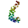
| Title | Crystal structure of a psychrophilic CCA-adding enzyme in complex with CMPcPP | |||||||||
 Components Components | CCA-adding enzyme CCA tRNA nucleotidyltransferase CCA tRNA nucleotidyltransferase | |||||||||
 Keywords Keywords | RNA BINDING PROTEIN / tRNA maturation / tRNA nucleotidyltransferase / psychrophilic enzyme | |||||||||
| Function / homology |  Function and homology information Function and homology informationRNA 3'-end processing / tRNA processing / nucleotidyltransferase activity / nucleotide binding / RNA binding / metal ion bindingSimilarity search - Function | |||||||||
| Biological species |  Planococcus halocryophilus (bacteria) Planococcus halocryophilus (bacteria) | |||||||||
| Method | X-RAY DIFFRACTION / SYNCHROTRON / MOLECULAR REPLACEMENT / Resolution: 2.2 Å | |||||||||
 Authors Authors | Rollet, K. / de Wijn, R. / Bluhm, A. / Hennig, O. / Betat, H. / Moerl, M. / Lorber, B. / Sauter, C. | |||||||||
| Funding support |  France, 2items France, 2items
| |||||||||
 Citation Citation | Journal: Comput Struct Biotechnol J / Year: 2021 Title: CCA-addition in the cold: Structural characterization of the psychrophilic CCA-adding enzyme from the permafrost bacterium Planococcus halocryophilus . Authors: de Wijn, R. / Rollet, K. / Ernst, F.G.M. / Wellner, K. / Betat, H. / Morl, M. / Sauter, C. | |||||||||
| History |
|
- Structure visualization
| Structure viewer | Molecule: MolmilJmol/JSmol |
|---|
- Downloads & links
-Download
| PDBx/mmCIF format | 7oqx.cif.gz | 106.5 KB | Display | PDBx/mmCIF format |
|---|---|---|---|---|
| PDB format | pdb7oqx.ent.gz | 73.2 KB | Display | PDB format |
| PDBx/mmJSON format | 7oqx.json.gz | Tree view | PDBx/mmJSON format | |
| Others |  Other downloads Other downloads |
-Validation report
| Arichive directory | https://data.pdbj.org/pub/pdb/validation_reports/oq/7oqxftp://data.pdbj.org/pub/pdb/validation_reports/oq/7oqx | HTTPS FTP |
|---|
-Related structure data
| Related structure data | 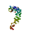 7otlC 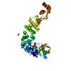 7otrC  6qxnS S: Starting model for refinement C: citing same article ( |
|---|---|
| Similar structure data |


-Links
 PDBj
PDBj- Assembly
| Deposited unit | 
| ||||||||||||
|---|---|---|---|---|---|---|---|---|---|---|---|---|---|
| 1 |
| ||||||||||||
| Unit cell |
|
-Components
-Protein , 1 types, 1 molecules A
| #1: Protein | CCA tRNA nucleotidyltransferase Mass: 48498.266 Da / Num. of mol.: 1 Source method: isolated from a genetically manipulated source Source: (gene. exp.) Planococcus halocryophilus (bacteria) / Gene: BBI08_05760 / Plasmid: pET-30b(+) / Details (production host): pET-30bEk/LIC / Production host: Escherichia coli BL21(DE3) (bacteria) / References: UniProt: A0A1C7DQ98 |
|---|
-Non-polymers , 5 types, 112 molecules 


| #2: Chemical | ChemComp-2TM /  Mass: 481.184 Da / Num. of mol.: 1 / Source method: obtained synthetically / Formula: C10H18N3O13P3 Mass: 481.184 Da / Num. of mol.: 1 / Source method: obtained synthetically / Formula: C10H18N3O13P3 | ||||||
|---|---|---|---|---|---|---|---|
| #3: Chemical | Phosphate Mass: 94.971 Da / Num. of mol.: 2 / Source method: obtained synthetically / Formula: PO4 Mass: 94.971 Da / Num. of mol.: 2 / Source method: obtained synthetically / Formula: PO4#4: Chemical | ChemComp-ACT / Acetate Mass: 59.044 Da / Num. of mol.: 9 / Source method: obtained synthetically / Formula: C2H3O2 Mass: 59.044 Da / Num. of mol.: 9 / Source method: obtained synthetically / Formula: C2H3O2#5: Chemical | ChemComp-GOL / | Glycerol Mass: 92.094 Da / Num. of mol.: 1 / Source method: obtained synthetically / Formula: C3H8O3 Mass: 92.094 Da / Num. of mol.: 1 / Source method: obtained synthetically / Formula: C3H8O3#6: Water | ChemComp-HOH / | WaterMass: 18.015 Da / Num. of mol.: 99 / Source method: isolated from a natural source / Formula: H2O |
-Details
| Has ligand of interest | N |
|---|
-Experimental details
-Experiment
| Experiment | Method: X-RAY DIFFRACTION / Number of used crystals: 1 |
|---|
- Sample preparation
| Crystal | Density Matthews: 3.66 Å3/Da / Density % sol: 66.37 % / Description: bipyramid |
|---|---|
| Crystal grow | Temperature: 293 K / Method: vapor diffusion, hanging drop Details: protein solution at 4.5 mg/mL in 50 mM Tris-HCl pH 7.5, 200 mM NaCl, 5 mM MgCl2; reservoir solution: 100 mM Sodium acetate; pH 4.5 1 M di-Ammonium hydrogen phosphate remarks : soaking 30 ...Details: protein solution at 4.5 mg/mL in 50 mM Tris-HCl pH 7.5, 200 mM NaCl, 5 mM MgCl2; reservoir solution: 100 mM Sodium acetate; pH 4.5 1 M di-Ammonium hydrogen phosphate remarks : soaking 30 seconds in a drop with 10 mM CMPcPP and reservoir solution + 20% glycerol PH range: 4.5 - 7.5 |
-Data collection
| Diffraction | Mean temperature: 100 K / Serial crystal experiment: N |
|---|---|
| Diffraction source | Source: SYNCHROTRON / Site: SOLEIL / Beamline: PROXIMA 1 / Wavelength: 0.9786 Å |
| Detector | Type: DECTRIS EIGER X 16M / Detector: PIXEL / Date: Oct 9, 2020 |
| Radiation | Protocol: SINGLE WAVELENGTH / Monochromatic (M) / Laue (L): M / Scattering type: x-ray |
| Radiation wavelength | Wavelength: 0.9786 Å / Relative weight: 1 |
| Reflection | Resolution: 2.2→50 Å / Num. obs: 37711 / % possible obs: 99.8 % / Redundancy: 25.7 % / Biso Wilson estimate: 50.85 Å2 / CC1/2: 0.99 / Rrim(I) all: 0.15 / Net I/av σ(I): 16.84 / Net I/σ(I): 16.84 |
| Reflection shell | Resolution: 2.2→2.34 Å / Redundancy: 27.1 % / Mean I/σ(I) obs: 1.98 / Num. unique obs: 5903 / CC1/2: 0.786 / Rrim(I) all: 1.616 / % possible all: 99.3 |
- Processing
| Software |
| ||||||||||||||||||||||||||||||||||||||||||||||||||||||||||||||||||||||||||||||||||||||||||||||||||
|---|---|---|---|---|---|---|---|---|---|---|---|---|---|---|---|---|---|---|---|---|---|---|---|---|---|---|---|---|---|---|---|---|---|---|---|---|---|---|---|---|---|---|---|---|---|---|---|---|---|---|---|---|---|---|---|---|---|---|---|---|---|---|---|---|---|---|---|---|---|---|---|---|---|---|---|---|---|---|---|---|---|---|---|---|---|---|---|---|---|---|---|---|---|---|---|---|---|---|---|
| Refinement | Method to determine structure: MOLECULAR REPLACEMENT Starting model: 6QXN Resolution: 2.2→46.74 Å / SU ML: 0.2954 / Cross valid method: FREE R-VALUE / σ(F): 1.34 / Phase error: 24.6168 Stereochemistry target values: GeoStd + Monomer Library + CDL v1.2
| ||||||||||||||||||||||||||||||||||||||||||||||||||||||||||||||||||||||||||||||||||||||||||||||||||
| Solvent computation | Shrinkage radii: 0.9 Å / VDW probe radii: 1.11 Å / Solvent model: FLAT BULK SOLVENT MODEL | ||||||||||||||||||||||||||||||||||||||||||||||||||||||||||||||||||||||||||||||||||||||||||||||||||
| Displacement parameters | Biso mean: 51.98 Å2 | ||||||||||||||||||||||||||||||||||||||||||||||||||||||||||||||||||||||||||||||||||||||||||||||||||
| Refinement step | Cycle: LAST / Resolution: 2.2→46.74 Å
| ||||||||||||||||||||||||||||||||||||||||||||||||||||||||||||||||||||||||||||||||||||||||||||||||||
| Refine LS restraints |
| ||||||||||||||||||||||||||||||||||||||||||||||||||||||||||||||||||||||||||||||||||||||||||||||||||
| LS refinement shell |
|
