 Movie
Movie Controller

[English] 日本語
 Yorodumi
- PDB-7odu: Natural killer cell receptor NKR-P1B from Rattus norvegicus in co... -
+ Open data

- Basic information
| Entry | Database: PDB / ID: 7odu | |||||||||
|---|---|---|---|---|---|---|---|---|---|---|
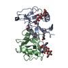
| Title | Natural killer cell receptor NKR-P1B from Rattus norvegicus in complex with its cognate ligand Clr-11 | |||||||||
 Components Components |
| |||||||||
 Keywords Keywords |  IMMUNE SYSTEM / RECEPTOR / CTLD FOLD / NATURAL KILLER CELL / PROTEIN COMPLEX IMMUNE SYSTEM / RECEPTOR / CTLD FOLD / NATURAL KILLER CELL / PROTEIN COMPLEX | |||||||||
| Function / homology |  Function and homology information Function and homology informationregulation of natural killer cell mediated cytotoxicity / natural killer cell lectin-like receptor binding / signaling receptor activity / carbohydrate binding / membrane => GO:0016020 / external side of plasma membrane / cell surface / plasma membraneSimilarity search - Function | |||||||||
| Biological species |  Rattus norvegicus (Norway rat) Rattus norvegicus (Norway rat) | |||||||||
| Method | X-RAY DIFFRACTION / SYNCHROTRON / MOLECULAR REPLACEMENT / Resolution: 3 Å | |||||||||
 Authors Authors | Skalova, T. / Blaha, J. / Kalouskova, B. / Skorepa, O. / Vanek, O. / Dohnalek, J. | |||||||||
| Funding support |  Czech Republic, 2items Czech Republic, 2items
| |||||||||
 Citation Citation | Journal: To Be Published Title: Natural killer cell receptor NKR-P1B from Rattus norvegicus in complex with its cognate ligand Clr-11 Authors: Skalova, T. / Vanek, O. / Kalouskova, B. / Skorepa, O. / Blaha, J. / Harlos, K. / Dohnalek, J. #1: Journal: Sci.Rep. / Year: 2019 Title: Production of recombinant soluble dimeric C-type lectin-like receptors of rat natural killer cells Authors: Vanek, O. / Celadova, P. / Skorepa, O. / Blaha, J. / Kalouskova, B. / Dvorska, A. / Polachova, E. / Pucholtova, H. / Kavan, D. / Pompach, P. / Hofbauerova, K. / Kopecky Jr, V. / Mesci, A. / ...Authors: Vanek, O. / Celadova, P. / Skorepa, O. / Blaha, J. / Kalouskova, B. / Dvorska, A. / Polachova, E. / Pucholtova, H. / Kavan, D. / Pompach, P. / Hofbauerova, K. / Kopecky Jr, V. / Mesci, A. / Voigt, S. / Carlyle, J.R. | |||||||||
| History |
|
- Structure visualization
| Structure viewer | Molecule: MolmilJmol/JSmol |
|---|
- Downloads & links
-Download
| PDBx/mmCIF format | 7odu.cif.gz | 94.7 KB | Display | PDBx/mmCIF format |
|---|---|---|---|---|
| PDB format | pdb7odu.ent.gz | 71 KB | Display | PDB format |
| PDBx/mmJSON format | 7odu.json.gz | Tree view | PDBx/mmJSON format | |
| Others |  Other downloads Other downloads |
-Validation report
| Arichive directory | https://data.pdbj.org/pub/pdb/validation_reports/od/7oduftp://data.pdbj.org/pub/pdb/validation_reports/od/7odu | HTTPS FTP |
|---|
-Related structure data
| Related structure data |  3ff8S S: Starting model for refinement |
|---|---|
| Similar structure data |
-Links
 PDBj
PDBj


- Assembly
| Deposited unit | 
| ||||||||||||||||||
|---|---|---|---|---|---|---|---|---|---|---|---|---|---|---|---|---|---|---|---|
| 1 | 
| ||||||||||||||||||
| 2 | 
| ||||||||||||||||||
| 3 | 
| ||||||||||||||||||
| Unit cell |
| ||||||||||||||||||
| Noncrystallographic symmetry (NCS) | NCS domain:
NCS domain segments: Component-ID: 0 / Ens-ID: 1 / Beg auth comp-ID: ILE / Beg label comp-ID: ILE / End auth comp-ID: ASN / End label comp-ID: ASN / Refine code: 0 / Auth seq-ID: 74 - 194 / Label seq-ID: 1 - 121
|
-Components
| #1: Protein | Mass: 15729.235 Da / Num. of mol.: 2 Source method: isolated from a genetically manipulated source Details: strain of rat: WAG / Source: (gene. exp.) Rattus norvegicus (Norway rat) / Strain: strain of rat: WAG / Gene: Clec2d11, Clrb, Ocil / Plasmid: pTW5sec / Details (production host): pTW5sec_C2 / Cell line (production host): HEK293S GnTI- / Organ (production host): KIDNEY / Production host:  Homo sapiens (human) / Tissue (production host): HUMAN EMBRYONIC KIDNEY / References: UniProt: Q0H8B9 Homo sapiens (human) / Tissue (production host): HUMAN EMBRYONIC KIDNEY / References: UniProt: Q0H8B9#2: Protein | | Mass: 16844.684 Da / Num. of mol.: 1 Source method: isolated from a genetically manipulated source Source: (gene. exp.) Rattus norvegicus (Norway rat) / Cell: NATURAL KILLER CELLS / Gene: Klrb1b, Nkrp1b / Plasmid: pYD5 / Details (production host): pYD5_S1 / Cell line (production host): HEK293S GnTI- / Organ (production host): KIDNEY / Production host: Homo sapiens (human) / Tissue (production host): HUMAN EMBRYONIC KIDNEY / References: UniProt: A4KWA1#3: Polysaccharide | / Mass: 424.401 Da / Num. of mol.: 2Source method: isolated from a genetically manipulated source #4: Sugar | N-Acetylglucosamine  Type: D-saccharide, beta linking / Mass: 221.208 Da / Num. of mol.: 3 Type: D-saccharide, beta linking / Mass: 221.208 Da / Num. of mol.: 3Source method: isolated from a genetically manipulated source Formula: C8H15NO6 #5: Chemical | ChemComp-ZN / |   Mass: 65.409 Da / Num. of mol.: 1 / Source method: obtained synthetically / Formula: Zn Mass: 65.409 Da / Num. of mol.: 1 / Source method: obtained synthetically / Formula: ZnHas ligand of interest | N | |
|---|
-Experimental details
-Experiment
| Experiment | Method: X-RAY DIFFRACTION / Number of used crystals: 1 |
|---|
- Sample preparation
| Crystal |
| ||||||||||||
|---|---|---|---|---|---|---|---|---|---|---|---|---|---|
| Crystal grow | Temperature: 293 K / Method: vapor diffusion, sitting drop / pH: 5 Details: 200 mM NaCl, 100 mM sodium acetate, 20 mM ZnCl2, 20% PEG 6000, pH 5.0 |
-Data collection
| Diffraction | Mean temperature: 100 K / Serial crystal experiment: N | |||||||||||||||||||||||||||
|---|---|---|---|---|---|---|---|---|---|---|---|---|---|---|---|---|---|---|---|---|---|---|---|---|---|---|---|---|
| Diffraction source | Source: SYNCHROTRON / Site: Diamond  / Beamline: I04 / Wavelength: 0.97625 Å / Beamline: I04 / Wavelength: 0.97625 Å | |||||||||||||||||||||||||||
| Detector | Type: DECTRIS PILATUS3 6M / Detector: PIXEL / Date: Apr 22, 2016 | |||||||||||||||||||||||||||
| Radiation | Protocol: SINGLE WAVELENGTH / Monochromatic (M) / Laue (L): M / Scattering type: x-ray | |||||||||||||||||||||||||||
| Radiation wavelength | Wavelength: 0.97625 Å / Relative weight: 1 | |||||||||||||||||||||||||||
| Reflection | Resolution: 3→49.77 Å / Num. obs: 16302 / % possible obs: 100 % / Redundancy: 8.4 % / Biso Wilson estimate: 91 Å2 / CC1/2: 0.999 / Rmerge(I) obs: 0.123 / Rpim(I) all: 0.045 / Rrim(I) all: 0.131 / Net I/σ(I): 10.6 | |||||||||||||||||||||||||||
| Reflection shell | Diffraction-ID: 1
|
- Processing
| Software |
| ||||||||||||||||||||||||||||||||||||||||||||||||||||||||||||
|---|---|---|---|---|---|---|---|---|---|---|---|---|---|---|---|---|---|---|---|---|---|---|---|---|---|---|---|---|---|---|---|---|---|---|---|---|---|---|---|---|---|---|---|---|---|---|---|---|---|---|---|---|---|---|---|---|---|---|---|---|---|
| Refinement | Method to determine structure: MOLECULAR REPLACEMENT Starting model: 3FF8 Resolution: 3→49.77 Å / Cor.coef. Fo:Fc: 0.945 / SU B: 16.169 / SU ML: 0.253 / Cross valid method: THROUGHOUT / σ(F): 0 / ESU R: 0.58 / Stereochemistry target values: MAXIMUM LIKELIHOOD Details: HYDROGENS HAVE BEEN ADDED IN THE RIDING POSITIONS; U VALUES WERE REFINED INDIVIDUALLY; ALL REFLECTIONS WERE USED IN THE LAST REFINEMENT CYCLE
| ||||||||||||||||||||||||||||||||||||||||||||||||||||||||||||
| Solvent computation | Ion probe radii: 0.8 Å / Shrinkage radii: 0.8 Å / VDW probe radii: 1.2 Å / Solvent model: MASK | ||||||||||||||||||||||||||||||||||||||||||||||||||||||||||||
| Displacement parameters | Biso max: 234.05 Å2 / Biso mean: 107.312 Å2 / Biso min: 70.92 Å2
| ||||||||||||||||||||||||||||||||||||||||||||||||||||||||||||
| Refinement step | Cycle: final / Resolution: 3→49.77 Å
| ||||||||||||||||||||||||||||||||||||||||||||||||||||||||||||
| Refine LS restraints |
| ||||||||||||||||||||||||||||||||||||||||||||||||||||||||||||
| Refine LS restraints NCS | Ens-ID: 1 / Number: 3880 / Refine-ID: X-RAY DIFFRACTION / Type: interatomic distance / Rms dev position: 0.13 Å / Weight position: 0.05
| ||||||||||||||||||||||||||||||||||||||||||||||||||||||||||||
| LS refinement shell | Resolution: 3→3.078 Å / Rfactor Rfree error: 0 / Total num. of bins used: 20
|
