 Movie
Movie Controller


+ Open data

- Basic information
| Entry | Database: PDB / ID: 7nh2 | |||||||||
|---|---|---|---|---|---|---|---|---|---|---|
| Title | Crystal structure of Toxoplasma CPSF4-YTH domain bound to m6A | |||||||||
 Components Components | Zinc finger (CCCH type) motif-containing protein | |||||||||
 Keywords Keywords |  RNA BINDING PROTEIN / YTH / Cleavage and Polyadenylation / RNA / m6A RNA BINDING PROTEIN / YTH / Cleavage and Polyadenylation / RNA / m6A | |||||||||
| Function / homology |  Function and homology information Function and homology informationN6-methyladenosine-containing RNA reader activity / regulation of alternative mRNA splicing, via spliceosome / mRNA splicing, via spliceosome / mRNA binding / nucleoplasm / metal ion bindingSimilarity search - Function | |||||||||
| Biological species |  Toxoplasma gondii (eukaryote) Toxoplasma gondii (eukaryote) | |||||||||
| Method | X-RAY DIFFRACTION / SYNCHROTRON / MOLECULAR REPLACEMENT / Resolution: 1.35 Å | |||||||||
 Authors Authors | Swale, C. / Bowler, M.W. | |||||||||
| Funding support |  France, European Union, 2items France, European Union, 2items
| |||||||||
 Citation Citation | Journal: Elife / Year: 2021 Title: A plant-like mechanism coupling m6A reading to polyadenylation safeguards transcriptome integrity and developmental gene partitioning in Toxoplasma . Authors: Farhat, D.C. / Bowler, M.W. / Communie, G. / Pontier, D. / Belmudes, L. / Mas, C. / Corrao, C. / Coute, Y. / Bougdour, A. / Lagrange, T. / Hakimi, M.A. / Swale, C. | |||||||||
| History |
|
- Structure visualization
| Structure viewer | Molecule: MolmilJmol/JSmol |
|---|
- Downloads & links
-Download
| PDBx/mmCIF format | 7nh2.cif.gz | 51.1 KB | Display | PDBx/mmCIF format |
|---|---|---|---|---|
| PDB format | pdb7nh2.ent.gz | 33.1 KB | Display | PDB format |
| PDBx/mmJSON format | 7nh2.json.gz | Tree view | PDBx/mmJSON format | |
| Others |  Other downloads Other downloads |
-Validation report
| Arichive directory | https://data.pdbj.org/pub/pdb/validation_reports/nh/7nh2ftp://data.pdbj.org/pub/pdb/validation_reports/nh/7nh2 | HTTPS FTP |
|---|
-Related structure data
| Related structure data | 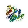 7ng2C 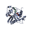 7njcC  4r3iS C: citing same article ( S: Starting model for refinement |
|---|---|
| Similar structure data |


-Links
 PDBj
PDBj
- Assembly
| Deposited unit | 
| ||||||||||||
|---|---|---|---|---|---|---|---|---|---|---|---|---|---|
| 1 |
| ||||||||||||
| Unit cell |
|
-Components
| #1: Protein | Mass: 18447.994 Da / Num. of mol.: 1 Source method: isolated from a genetically manipulated source Source: (gene. exp.) Toxoplasma gondii (strain ATCC 50611 / Me49) (eukaryote)Strain: ATCC 50611 / Me49 / Gene: TGME49_201200 / Production host:  Escherichia coli (E. coli) / Strain (production host): BL21-RIL / References: UniProt: S8F6K2 Escherichia coli (E. coli) / Strain (production host): BL21-RIL / References: UniProt: S8F6K2 |
|---|---|
| #2: Chemical | ChemComp-PG4 / Polyethylene glycol  Mass: 194.226 Da / Num. of mol.: 1 / Source method: obtained synthetically / Formula: C8H18O5 / Comment: precipitant*YM Mass: 194.226 Da / Num. of mol.: 1 / Source method: obtained synthetically / Formula: C8H18O5 / Comment: precipitant*YM |
| #3: Chemical | ChemComp-OYK / ~{  Mass: 163.180 Da / Num. of mol.: 1 / Source method: obtained synthetically / Formula: C7H9N5 / Feature type: SUBJECT OF INVESTIGATION Mass: 163.180 Da / Num. of mol.: 1 / Source method: obtained synthetically / Formula: C7H9N5 / Feature type: SUBJECT OF INVESTIGATION |
| #4: Water | ChemComp-HOH / Water Mass: 18.015 Da / Num. of mol.: 148 / Source method: isolated from a natural source / Formula: H2O Mass: 18.015 Da / Num. of mol.: 148 / Source method: isolated from a natural source / Formula: H2O |
| Has ligand of interest | Y |
-Experimental details
-Experiment
| Experiment | Method: X-RAY DIFFRACTION / Number of used crystals: 1 |
|---|
- Sample preparation
| Crystal | Density Matthews: 2.06 Å3/Da / Density % sol: 40.25 % |
|---|---|
| Crystal grow | Temperature: 293 K / Method: vapor diffusion, sitting drop / pH: 4.5 Details: 50% PEG 400, 0.1M Sodium acetate pH 4.5 and 0.2M LiSO4 |
-Data collection
| Diffraction | Mean temperature: 100 K / Serial crystal experiment: N |
|---|---|
| Diffraction source | Source: SYNCHROTRON / Site: ESRF / Beamline: MASSIF-1 / Wavelength: 0.966 Å |
| Detector | Type: DECTRIS PILATUS3 2M / Detector: PIXEL / Date: Sep 26, 2018 |
| Radiation | Protocol: SINGLE WAVELENGTH / Monochromatic (M) / Laue (L): M / Scattering type: x-ray |
| Radiation wavelength | Wavelength: 0.966 Å / Relative weight: 1 |
| Reflection | Resolution: 1.349→30.76 Å / Num. obs: 30036 / % possible obs: 92.64 % / Redundancy: 1.6 % / Biso Wilson estimate: 9.18 Å2 / CC1/2: 0.956 / Net I/σ(I): 6.6 |
| Reflection shell | Resolution: 1.349→1.397 Å / Redundancy: 1.7 % / Mean I/σ(I) obs: 2.1 / Num. unique obs: 2887 / CC1/2: 0.496 / % possible all: 88.38 |
- Processing
| Software |
| ||||||||||||||||||||||||||||||||||||||||||||||||||||||||||||||||||||||||||||||||||||
|---|---|---|---|---|---|---|---|---|---|---|---|---|---|---|---|---|---|---|---|---|---|---|---|---|---|---|---|---|---|---|---|---|---|---|---|---|---|---|---|---|---|---|---|---|---|---|---|---|---|---|---|---|---|---|---|---|---|---|---|---|---|---|---|---|---|---|---|---|---|---|---|---|---|---|---|---|---|---|---|---|---|---|---|---|---|
| Refinement | Method to determine structure: MOLECULAR REPLACEMENT Starting model: 4r3i Resolution: 1.35→30.76 Å / SU ML: 0.1393 / Cross valid method: FREE R-VALUE / σ(F): 1.96 / Phase error: 20.9977 Stereochemistry target values: GeoStd + Monomer Library + CDL v1.2
| ||||||||||||||||||||||||||||||||||||||||||||||||||||||||||||||||||||||||||||||||||||
| Solvent computation | Shrinkage radii: 0.9 Å / VDW probe radii: 1.11 Å / Solvent model: FLAT BULK SOLVENT MODEL | ||||||||||||||||||||||||||||||||||||||||||||||||||||||||||||||||||||||||||||||||||||
| Displacement parameters | Biso mean: 13.21 Å2 | ||||||||||||||||||||||||||||||||||||||||||||||||||||||||||||||||||||||||||||||||||||
| Refinement step | Cycle: LAST / Resolution: 1.35→30.76 Å
| ||||||||||||||||||||||||||||||||||||||||||||||||||||||||||||||||||||||||||||||||||||
| Refine LS restraints |
| ||||||||||||||||||||||||||||||||||||||||||||||||||||||||||||||||||||||||||||||||||||
| LS refinement shell |
|
