 Movie
Movie Controller


+ Open data

- Basic information
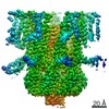
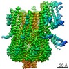
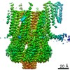
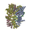
| Entry | Database: PDB / ID: 7n5e | |||||||||||||||
|---|---|---|---|---|---|---|---|---|---|---|---|---|---|---|---|---|
| Title | Structure of Mechanosensitive Ion Channel Flycatcher1 in GDN | |||||||||||||||
 Components Components | Mechanosensitive ion channel Flycatcher1 | |||||||||||||||
 Keywords Keywords |  MEMBRANE PROTEIN / mechanically activated ion channel MEMBRANE PROTEIN / mechanically activated ion channel | |||||||||||||||
| Function / homology | PALMITIC ACID Function and homology information Function and homology information | |||||||||||||||
| Biological species |  Dionaea muscipula (Venus flytrap) Dionaea muscipula (Venus flytrap) | |||||||||||||||
| Method | ELECTRON MICROSCOPY / single particle reconstruction / cryo EM / Resolution: 2.8 Å | |||||||||||||||
 Authors Authors | Jojoa-Cruz, S. / Saotome, K. / Lee, W.H. / Patapoutian, A. / Ward, A.B. | |||||||||||||||
| Funding support |  United States, United States,  United Kingdom, 4items United Kingdom, 4items
| |||||||||||||||
 Citation Citation | Journal: Nat Commun / Year: 2022 Title: Structural insights into the Venus flytrap mechanosensitive ion channel Flycatcher1. Authors: Sebastian Jojoa-Cruz / Kei Saotome / Che Chun Alex Tsui / Wen-Hsin Lee / Mark S P Sansom / Swetha E Murthy / Ardem Patapoutian / Andrew B Ward / Abstract: Flycatcher1 (FLYC1), a MscS homolog, has recently been identified as a candidate mechanosensitive (MS) ion channel involved in Venus flytrap prey recognition. FLYC1 is a larger protein and its ...Flycatcher1 (FLYC1), a MscS homolog, has recently been identified as a candidate mechanosensitive (MS) ion channel involved in Venus flytrap prey recognition. FLYC1 is a larger protein and its sequence diverges from previously studied MscS homologs, suggesting it has unique structural features that contribute to its function. Here, we characterize FLYC1 by cryo-electron microscopy, molecular dynamics simulations, and electrophysiology. Akin to bacterial MscS and plant MSL1 channels, we find that FLYC1 central core includes side portals in the cytoplasmic cage that regulate ion preference and conduction, by identifying critical residues that modulate channel conductance. Topologically unique cytoplasmic flanking regions can adopt 'up' or 'down' conformations, making the channel asymmetric. Disruption of an up conformation-specific interaction severely delays channel deactivation by 40-fold likely due to stabilization of the channel open state. Our results illustrate novel structural features and likely conformational transitions that regulate mechano-gating of FLYC1. | |||||||||||||||
| History |
|
- Structure visualization
| Movie |
Movie viewer |
|---|---|
| Structure viewer | Molecule: MolmilJmol/JSmol |
- Downloads & links
-Download
| PDBx/mmCIF format | 7n5e.cif.gz | 465.3 KB | Display | PDBx/mmCIF format |
|---|---|---|---|---|
| PDB format | pdb7n5e.ent.gz | 381.8 KB | Display | PDB format |
| PDBx/mmJSON format | 7n5e.json.gz | Tree view | PDBx/mmJSON format | |
| Others |  Other downloads Other downloads |
-Validation report
| Arichive directory | https://data.pdbj.org/pub/pdb/validation_reports/n5/7n5eftp://data.pdbj.org/pub/pdb/validation_reports/n5/7n5e | HTTPS FTP |
|---|
-Related structure data
| Related structure data | 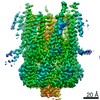 24187MC 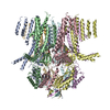 7n5dC 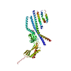 7n5fC 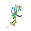 7n5gC C: citing same article ( M: map data used to model this data |
|---|---|
| Similar structure data | |
| EM raw data | EMPIAR-10740 (Title: Cryo-EM of Mechanosensitive Ion Channel Flycatcher1 in GDN Data size: 5.7 TB Data #1: Unaligned multi-frame micrographs of FLYC1 in GDN, dataset 1 [micrographs - multiframe] Data #2: Unaligned multi-frame micrographs of FLYC1 in GDN, dataset 2 [micrographs - multiframe] Data #3: Gain references for dataset 1 and dataset 2 [micrographs - single frame]) |
| Experimental dataset #1 | Data reference: 10.6019/EMPIAR-10740 / Data set type: EMPIAR |


-Links
 PDBj
PDBj


- Assembly
| Deposited unit | 
|
|---|---|
| 1 |
|
-Components
| #1: Protein | Mass: 86814.523 Da / Num. of mol.: 7 Source method: isolated from a genetically manipulated source Source: (gene. exp.) Dionaea muscipula (Venus flytrap) / Tissue: Trigger hair / Organ: Trap / Plasmid: pEG / Cell line (production host): HEK293F / Production host:  Homo sapiens (human) Homo sapiens (human)#2: Chemical | ChemComp-PLM / Palmitic acid  Mass: 256.424 Da / Num. of mol.: 7 / Source method: obtained synthetically / Formula: C16H32O2 Mass: 256.424 Da / Num. of mol.: 7 / Source method: obtained synthetically / Formula: C16H32O2Has ligand of interest | N | Sequence details | The last 10 residues (GSGSLEVLFQ | |
|---|
-Experimental details
-Experiment
| Experiment | Method: ELECTRON MICROSCOPY |
|---|---|
| EM experiment | Aggregation state: PARTICLE / 3D reconstruction method: single particle reconstruction |
- Sample preparation
| Component | Name: FLYC1 / Type: ORGANELLE OR CELLULAR COMPONENT / Entity ID: #1 / Source: RECOMBINANT | ||||||||||||||||||||||||||||||||||||
|---|---|---|---|---|---|---|---|---|---|---|---|---|---|---|---|---|---|---|---|---|---|---|---|---|---|---|---|---|---|---|---|---|---|---|---|---|---|
| Molecular weight | Value: 0.607 MDa / Experimental value: NO | ||||||||||||||||||||||||||||||||||||
| Source (natural) | Organism: Dionaea muscipula (Venus flytrap) / Organ: Trap / Tissue: Trigger hair | ||||||||||||||||||||||||||||||||||||
| Source (recombinant) | Organism: Homo sapiens (human) / Cell: HEK293F / Plasmid: pEG | ||||||||||||||||||||||||||||||||||||
| Buffer solution | pH: 8 | ||||||||||||||||||||||||||||||||||||
| Buffer component |
| ||||||||||||||||||||||||||||||||||||
| Specimen | Conc.: 7 mg/ml / Embedding applied: NO / Shadowing applied: NO / Staining applied: NO / Vitrification applied: YES | ||||||||||||||||||||||||||||||||||||
| Vitrification | Instrument: FEI VITROBOT MARK IV / Cryogen name: ETHANE / Humidity: 100 % / Chamber temperature: 283 K |
- Electron microscopy imaging
| Experimental equipment |  Model: Titan Krios / Image courtesy: FEI Company |
|---|---|
| Microscopy | Model: FEI TITAN KRIOS |
| Electron gun | Electron source: FIELD EMISSION GUN / Accelerating voltage: 300 kV / Illumination mode: FLOOD BEAM |
| Electron lens | Mode: BRIGHT FIELDBright-field microscopy / Nominal magnification: 29000 X / Nominal defocus max: 2200 nm / Nominal defocus min: 700 nm / Cs: 2.7 mm / C2 aperture diameter: 70 µm / Alignment procedure: COMA FREE |
| Specimen holder | Cryogen: NITROGEN / Specimen holder model: FEI TITAN KRIOS AUTOGRID HOLDER |
| Image recording | Electron dose: 50 e/Å2 / Detector mode: COUNTING / Film or detector model: GATAN K2 SUMMIT (4k x 4k) |
| Image scans | Width: 3710 / Height: 3838 / Movie frames/image: 43 |
- Processing
| EM software |
| |||||||||||||||||||||||||||||||||||||||||||||||||||||||||||||||||
|---|---|---|---|---|---|---|---|---|---|---|---|---|---|---|---|---|---|---|---|---|---|---|---|---|---|---|---|---|---|---|---|---|---|---|---|---|---|---|---|---|---|---|---|---|---|---|---|---|---|---|---|---|---|---|---|---|---|---|---|---|---|---|---|---|---|---|
| CTF correction | Type: PHASE FLIPPING AND AMPLITUDE CORRECTION | |||||||||||||||||||||||||||||||||||||||||||||||||||||||||||||||||
| Particle selection | Num. of particles selected: 1100000 | |||||||||||||||||||||||||||||||||||||||||||||||||||||||||||||||||
| Symmetry | Point symmetry: C1 (asymmetric) | |||||||||||||||||||||||||||||||||||||||||||||||||||||||||||||||||
| 3D reconstruction | Resolution: 2.8 Å / Resolution method: FSC 0.143 CUT-OFF / Num. of particles: 255359 / Num. of class averages: 1 / Symmetry type: POINT | |||||||||||||||||||||||||||||||||||||||||||||||||||||||||||||||||
| Atomic model building | Protocol: AB INITIO MODEL / Space: REAL |
