 Movie
Movie Controller


+ Open data

- Basic information
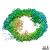
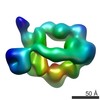
| Entry | Database: PDB / ID: 7mwe | ||||||||||||||||||
|---|---|---|---|---|---|---|---|---|---|---|---|---|---|---|---|---|---|---|---|
| Title | HUWE1 in map with focus on WWE | ||||||||||||||||||
 Components Components | E3 ubiquitin-protein ligase HUWE1 | ||||||||||||||||||
 Keywords Keywords |  TRANSFERASE / Ubiquitin / Quality Control / E3 ligase / protein degradation TRANSFERASE / Ubiquitin / Quality Control / E3 ligase / protein degradation | ||||||||||||||||||
| Function / homology |  Function and homology information Function and homology informationnegative regulation of mitochondrial fusion / positive regulation of mitophagy in response to mitochondrial depolarization / histone ubiquitin ligase activity / HECT-type E3 ubiquitin transferase / positive regulation of protein targeting to mitochondrion / Golgi organization / protein monoubiquitination / positive regulation of protein ubiquitination / circadian regulation of gene expression / base-excision repair ...negative regulation of mitochondrial fusion / positive regulation of mitophagy in response to mitochondrial depolarization / histone ubiquitin ligase activity / HECT-type E3 ubiquitin transferase / positive regulation of protein targeting to mitochondrion / Golgi organization / protein monoubiquitination / positive regulation of protein ubiquitination / circadian regulation of gene expression / base-excision repair / protein polyubiquitination / ubiquitin-protein transferase activity / ubiquitin protein ligase activity / Antigen processing: Ubiquitination & Proteasome degradation / secretory granule lumen / ficolin-1-rich granule lumen / membrane fusion / cell differentiation / Golgi membrane / Neutrophil degranulation / mitochondrion / DNA binding / RNA binding / extracellular exosome / extracellular region / nucleoplasm / membrane / nucleus / cytosol / cytoplasmSimilarity search - Function | ||||||||||||||||||
| Biological species |  Homo sapiens (human) Homo sapiens (human) | ||||||||||||||||||
| Method | ELECTRON MICROSCOPY / single particle reconstruction / cryo EM / Resolution: 3.4 Å | ||||||||||||||||||
 Authors Authors | Hunkeler, M. / Fischer, E.S. | ||||||||||||||||||
| Funding support |  United States, United States,  Switzerland, 5items Switzerland, 5items
| ||||||||||||||||||
 Citation Citation | Journal: Mol Cell / Year: 2021 Title: Solenoid architecture of HUWE1 contributes to ligase activity and substrate recognition. Authors: Moritz Hunkeler / Cyrus Y Jin / Michelle W Ma / Julie K Monda / Daan Overwijn / Eric J Bennett / Eric S Fischer / Abstract: HECT ubiquitin ligases play essential roles in metazoan development and physiology. The HECT ligase HUWE1 is central to the cellular stress response by mediating degradation of key death or survival ...HECT ubiquitin ligases play essential roles in metazoan development and physiology. The HECT ligase HUWE1 is central to the cellular stress response by mediating degradation of key death or survival factors, including Mcl1, p53, DDIT4, and Myc. Although mutations in HUWE1 and related HECT ligases are widely implicated in human disease, our molecular understanding remains limited. Here we present a comprehensive investigation of full-length HUWE1, deepening our understanding of this class of enzymes. The N-terminal ∼3,900 amino acids of HUWE1 are indispensable for proper ligase function, and our cryo-EM structures of HUWE1 offer a complete molecular picture of this large HECT ubiquitin ligase. HUWE1 forms an alpha solenoid-shaped assembly with a central pore decorated with protein interaction modules. Structures of HUWE1 variants linked to neurodevelopmental disorders as well as of HUWE1 bound to a model substrate link the functions of this essential enzyme to its three-dimensional organization. | ||||||||||||||||||
| History |
|
- Structure visualization
| Movie |
Movie viewer |
|---|---|
| Structure viewer | Molecule: MolmilJmol/JSmol |
- Downloads & links
-Download
| PDBx/mmCIF format | 7mwe.cif.gz | 858.5 KB | Display | PDBx/mmCIF format |
|---|---|---|---|---|
| PDB format | pdb7mwe.ent.gz | 673.8 KB | Display | PDB format |
| PDBx/mmJSON format | 7mwe.json.gz | Tree view | PDBx/mmJSON format | |
| Others |  Other downloads Other downloads |
-Validation report
| Arichive directory | https://data.pdbj.org/pub/pdb/validation_reports/mw/7mweftp://data.pdbj.org/pub/pdb/validation_reports/mw/7mwe | HTTPS FTP |
|---|
-Related structure data
| Related structure data |  22429MC 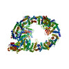 7jq9C 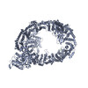 7mopC 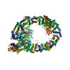 7mwdC  7mwfC C: citing same article ( M: map data used to model this data |
|---|---|
| Similar structure data |


-Links
 PDBj
PDBj

- Assembly
| Deposited unit | 
|
|---|---|
| 1 |
|
-Components
| #1: Protein | Mass: 486409.531 Da / Num. of mol.: 1 Source method: isolated from a genetically manipulated source Source: (gene. exp.) Homo sapiens (human) / Gene: HUWE1, KIAA0312, KIAA1578, UREB1, HSPC272 / Plasmid: pDEST / Cell line (production host): Expi293 / Production host: Homo sapiens (human)References: UniProt: Q7Z6Z7, HECT-type E3 ubiquitin transferase |
|---|
-Experimental details
-Experiment
| Experiment | Method: ELECTRON MICROSCOPY |
|---|---|
| EM experiment | Aggregation state: PARTICLE / 3D reconstruction method: single particle reconstruction |
- Sample preparation
| Component | Name: E3 ubiquitin-protein ligase HUWE1 / Type: ORGANELLE OR CELLULAR COMPONENT / Details: full length, crosslinked with BS3 / Entity ID: all / Source: RECOMBINANT | |||||||||||||||
|---|---|---|---|---|---|---|---|---|---|---|---|---|---|---|---|---|
| Molecular weight | Value: 0.48 MDa / Experimental value: NO | |||||||||||||||
| Source (natural) | Organism: Homo sapiens (human) | |||||||||||||||
| Source (recombinant) | Organism: Homo sapiens (human) / Strain: Expi293 / Plasmid: pDEST | |||||||||||||||
| Buffer solution | pH: 7.4 | |||||||||||||||
| Buffer component |
| |||||||||||||||
| Specimen | Conc.: 0.9 mg/ml / Embedding applied: NO / Shadowing applied: NO / Staining applied: NO / Vitrification applied: YES / Details: Sample crosslinked with BS3. Monodisperse. | |||||||||||||||
| Specimen support | Grid material: COPPER / Grid mesh size: 300 divisions/in. / Grid type: Quantifoil R1.2/1.3 | |||||||||||||||
| Vitrification | Instrument: LEICA EM GP / Cryogen name: ETHANE / Humidity: 95 % / Chamber temperature: 283 K Details: CHAPSO detergent added to final conc. of 0.8 mM. Sample applied twice. |
- Electron microscopy imaging
| Experimental equipment |  Model: Titan Krios / Image courtesy: FEI Company |
|---|---|
| Microscopy | Model: FEI TITAN KRIOS Details: Data collection in counting mode, using multi-shot scheme (4 holes per stage position, 3 movies per hole) |
| Electron gun | Electron source: FIELD EMISSION GUN / Accelerating voltage: 300 kV / Illumination mode: FLOOD BEAM |
| Electron lens | Mode: BRIGHT FIELDBright-field microscopy / Nominal magnification: 105000 X / Nominal defocus max: -2500 nm / Nominal defocus min: -800 nm / Cs: 2.7 mm / C2 aperture diameter: 50 µm / Alignment procedure: COMA FREE |
| Specimen holder | Cryogen: NITROGEN / Specimen holder model: FEI TITAN KRIOS AUTOGRID HOLDER |
| Image recording | Average exposure time: 2.4 sec. / Electron dose: 45.68 e/Å2 / Film or detector model: GATAN K3 BIOQUANTUM (6k x 4k) / Num. of grids imaged: 1 / Num. of real images: 10390 |
| Image scans | Width: 5760 / Height: 4092 |
- Processing
| Software |
| |||||||||||||||||||||||||||||||||||||||
|---|---|---|---|---|---|---|---|---|---|---|---|---|---|---|---|---|---|---|---|---|---|---|---|---|---|---|---|---|---|---|---|---|---|---|---|---|---|---|---|---|
| EM software |
| |||||||||||||||||||||||||||||||||||||||
| CTF correction | Details: standard correction in Relion / Type: PHASE FLIPPING AND AMPLITUDE CORRECTION | |||||||||||||||||||||||||||||||||||||||
| Particle selection | Num. of particles selected: 2110785 | |||||||||||||||||||||||||||||||||||||||
| Symmetry | Point symmetry: C1 (asymmetric) | |||||||||||||||||||||||||||||||||||||||
| 3D reconstruction | Resolution: 3.4 Å / Resolution method: FSC 0.143 CUT-OFF / Num. of particles: 85184 / Algorithm: BACK PROJECTION / Details: as implemented in Relion / Num. of class averages: 1 / Symmetry type: POINT | |||||||||||||||||||||||||||||||||||||||
| Atomic model building | B value: 63.97 / Protocol: AB INITIO MODEL / Space: REAL / Target criteria: CC | |||||||||||||||||||||||||||||||||||||||
| Refinement | Cross valid method: NONE Stereochemistry target values: GeoStd + Monomer Library + CDL v1.2 | |||||||||||||||||||||||||||||||||||||||
| Displacement parameters | Biso mean: 63.97 Å2 | |||||||||||||||||||||||||||||||||||||||
| Refine LS restraints |
|
