 Movie
Movie Controller

[English] 日本語
 Yorodumi
- PDB-7mt1: Crystal structure of Human Platelet-activating factor acetylhydro... -
+ Open data

- Basic information
| Entry | Database: PDB / ID: 7mt1 | ||||||
|---|---|---|---|---|---|---|---|
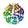
| Title | Crystal structure of Human Platelet-activating factor acetylhydrolase IB subunit beta (PAFAH1B1) | ||||||
 Components Components | Platelet-activating factor acetylhydrolase IB subunit beta | ||||||
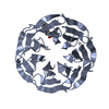
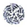
 Keywords Keywords |  PROTEIN BINDING / PAFAH1B1 / WD40 / WD-repeat protein / Structural Genomics / Structural Genomics Consortium / SGC PROTEIN BINDING / PAFAH1B1 / WD40 / WD-repeat protein / Structural Genomics / Structural Genomics Consortium / SGC | ||||||
| Function / homology |  Function and homology information Function and homology informationcorpus callosum morphogenesis / establishment of planar polarity of embryonic epithelium / microtubule cytoskeleton organization involved in establishment of planar polarity / ameboidal-type cell migration / interneuron migration / 1-alkyl-2-acetylglycerophosphocholine esterase complex / maintenance of centrosome location / microtubule sliding / platelet activating factor metabolic process / acrosome assembly ...corpus callosum morphogenesis / establishment of planar polarity of embryonic epithelium / microtubule cytoskeleton organization involved in establishment of planar polarity / ameboidal-type cell migration / interneuron migration / 1-alkyl-2-acetylglycerophosphocholine esterase complex / maintenance of centrosome location / microtubule sliding / platelet activating factor metabolic process / acrosome assembly / radial glia-guided pyramidal neuron migration / microtubule organizing center organization / cerebral cortex neuron differentiation / central region of growth cone / positive regulation of embryonic development / reelin-mediated signaling pathway / establishment of centrosome localization / positive regulation of cytokine-mediated signaling pathway / cortical microtubule organization / astral microtubule / layer formation in cerebral cortex / nuclear membrane disassembly / auditory receptor cell development / positive regulation of dendritic spine morphogenesis / vesicle transport along microtubule / stem cell division / stereocilium / myeloid leukocyte migration / COPI-independent Golgi-to-ER retrograde traffic / microtubule plus-end binding / retrograde axonal transport / negative regulation of JNK cascade / brain morphogenesis / motile cilium / nuclear migration / osteoclast development / microtubule associated complex / kinesin complex / dynein intermediate chain binding / dynein complex binding / cochlea development / transmission of nerve impulse / cell leading edge / germ cell development / dynactin binding / establishment of mitotic spindle orientation / phospholipase binding / neuromuscular process controlling balance / neuroblast proliferation / protein secretion / positive regulation of axon extension / Amplification of signal from unattached kinetochores via a MAD2 inhibitory signal / cytoplasmic microtubule / microtubule-based process / lipid catabolic process / regulation of microtubule cytoskeleton organization / Mitotic Prometaphase / EML4 and NUDC in mitotic spindle formation / axon cytoplasm / JNK cascade / Loss of Nlp from mitotic centrosomes / Loss of proteins required for interphase microtubule organization from the centrosome / Recruitment of mitotic centrosome proteins and complexes / Resolution of Sister Chromatid Cohesion / Recruitment of NuMA to mitotic centrosomes / Anchoring of the basal body to the plasma membrane / positive regulation of mitotic cell cycle / adult locomotory behavior / AURKA Activation by TPX2 / RHO GTPases Activate Formins / hippocampus development / phosphoprotein binding / neuron migration / modulation of chemical synaptic transmission / Schaffer collateral - CA1 synapse / kinetochore / microtubule cytoskeleton organization / cerebral cortex development / Separation of Sister Chromatids / Regulation of PLK1 Activity at G2/M Transition / negative regulation of neuron projection development / nuclear envelope / heparin binding / cell cortex / actin cytoskeleton organization / chemical synaptic transmission / microtubule binding / nuclear membrane / learning or memory / neuron projection / protein heterodimerization activity / centrosome / neuronal cell body / glutamatergic synapse / perinuclear region of cytoplasm / extracellular exosome / identical protein binding / cytosolSimilarity search - Function | ||||||
| Biological species |  Homo sapiens (human) Homo sapiens (human) | ||||||
| Method | X-RAY DIFFRACTION / SYNCHROTRON / MOLECULAR REPLACEMENT / Resolution: 1.3 Å | ||||||
 Authors Authors | Hutchinson, A. / Seitova, A. / Dong, A. / Loppnau, P. / Edwards, A.M. / Arrowsmith, C.H. / Halabelian, L. / Structural Genomics Consortium (SGC) | ||||||
 Citation Citation | Journal: To Be Published Title: Crystal structure of Human Platelet-activating factor acetylhydrolase IB subunit beta (PAFAH1B1) Authors: Hutchinson, A. / Seitova, A. / Dong, A. / Loppnau, P. / Edwards, A.M. / Arrowsmith, C.H. / Halabelian, L. / Structural Genomics Consortium (SGC) | ||||||
| History |
|
- Structure visualization
| Structure viewer | Molecule: MolmilJmol/JSmol |
|---|
- Downloads & links
-Download
| PDBx/mmCIF format | 7mt1.cif.gz | 86.4 KB | Display | PDBx/mmCIF format |
|---|---|---|---|---|
| PDB format | pdb7mt1.ent.gz | 61.1 KB | Display | PDB format |
| PDBx/mmJSON format | 7mt1.json.gz | Tree view | PDBx/mmJSON format | |
| Others |  Other downloads Other downloads |
-Validation report
| Arichive directory | https://data.pdbj.org/pub/pdb/validation_reports/mt/7mt1ftp://data.pdbj.org/pub/pdb/validation_reports/mt/7mt1 | HTTPS FTP |
|---|
-Related structure data
| Related structure data | 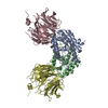 1vyhS S: Starting model for refinement |
|---|---|
| Similar structure data |


-Links
 PDBj
PDBj


- Assembly
| Deposited unit | 
| ||||||||
|---|---|---|---|---|---|---|---|---|---|
| 1 |
| ||||||||
| Unit cell |
|
-Components
| #1: Protein | Mass: 39014.176 Da / Num. of mol.: 1 Source method: isolated from a genetically manipulated source Source: (gene. exp.) Homo sapiens (human) / Gene: PAFAH1B1, LIS1, MDCR, MDS, PAFAHA / Plasmid: pFBOH-MHL / Cell line (production host): Sf9 / Production host:   Spodoptera frugiperda (fall armyworm) / References: UniProt: P43034 Spodoptera frugiperda (fall armyworm) / References: UniProt: P43034 | ||||
|---|---|---|---|---|---|
| #2: Chemical | ChemComp-GOL / Glycerol  Mass: 92.094 Da / Num. of mol.: 1 / Source method: obtained synthetically / Formula: C3H8O3 Mass: 92.094 Da / Num. of mol.: 1 / Source method: obtained synthetically / Formula: C3H8O3 | ||||
| #3: Chemical | ChemComp-UNX /   Num. of mol.: 4 / Source method: obtained synthetically Num. of mol.: 4 / Source method: obtained synthetically#4: Water | ChemComp-HOH / | Water Mass: 18.015 Da / Num. of mol.: 251 / Source method: isolated from a natural source / Formula: H2O Mass: 18.015 Da / Num. of mol.: 251 / Source method: isolated from a natural source / Formula: H2OHas ligand of interest | N | |
-Experimental details
-Experiment
| Experiment | Method: X-RAY DIFFRACTION / Number of used crystals: 1 |
|---|
- Sample preparation
| Crystal | Density Matthews: 2.2 Å3/Da / Density % sol: 44.13 % / Mosaicity: 0.09 ° |
|---|---|
| Crystal grow | Temperature: 295 K / Method: vapor diffusion, sitting drop / pH: 8.5 / Details: 18% PEG8K, 0.1M Tris pH 8.5 |
-Data collection
| Diffraction | Mean temperature: 100 K / Serial crystal experiment: N | ||||||||||||||||||||||||||||||
|---|---|---|---|---|---|---|---|---|---|---|---|---|---|---|---|---|---|---|---|---|---|---|---|---|---|---|---|---|---|---|---|
| Diffraction source | Source: SYNCHROTRON / Site: APS  / Beamline: 24-ID-E / Wavelength: 0.97918 Å / Beamline: 24-ID-E / Wavelength: 0.97918 Å | ||||||||||||||||||||||||||||||
| Detector | Type: DECTRIS EIGER2 X 16M / Detector: PIXEL / Date: Apr 11, 2021 | ||||||||||||||||||||||||||||||
| Radiation | Protocol: SINGLE WAVELENGTH / Monochromatic (M) / Laue (L): M / Scattering type: x-ray | ||||||||||||||||||||||||||||||
| Radiation wavelength | Wavelength: 0.97918 Å / Relative weight: 1 | ||||||||||||||||||||||||||||||
| Reflection | Resolution: 1.3→36.04 Å / Num. obs: 82396 / % possible obs: 99.2 % / Redundancy: 5.6 % / CC1/2: 1 / Rmerge(I) obs: 0.034 / Rpim(I) all: 0.015 / Rrim(I) all: 0.037 / Net I/σ(I): 22.5 | ||||||||||||||||||||||||||||||
| Reflection shell | Diffraction-ID: 1
|
- Processing
| Software |
| ||||||||||||||||||||||||||||||||||||||||||||||||||||||||||||
|---|---|---|---|---|---|---|---|---|---|---|---|---|---|---|---|---|---|---|---|---|---|---|---|---|---|---|---|---|---|---|---|---|---|---|---|---|---|---|---|---|---|---|---|---|---|---|---|---|---|---|---|---|---|---|---|---|---|---|---|---|---|
| Refinement | Method to determine structure: MOLECULAR REPLACEMENT Starting model: 1vyh Resolution: 1.3→36.03 Å / Cor.coef. Fo:Fc: 0.97 / Cor.coef. Fo:Fc free: 0.962 / SU B: 0.728 / SU ML: 0.031 / Cross valid method: THROUGHOUT / σ(F): 0 / ESU R: 0.048 / ESU R Free: 0.049 / Stereochemistry target values: MAXIMUM LIKELIHOOD Details: HYDROGENS HAVE BEEN ADDED IN THE RIDING POSITIONS U VALUES : REFINED INDIVIDUALLY
| ||||||||||||||||||||||||||||||||||||||||||||||||||||||||||||
| Solvent computation | Ion probe radii: 0.8 Å / Shrinkage radii: 0.8 Å / VDW probe radii: 1.2 Å / Solvent model: MASK | ||||||||||||||||||||||||||||||||||||||||||||||||||||||||||||
| Displacement parameters | Biso max: 73.33 Å2 / Biso mean: 15.789 Å2 / Biso min: 8.26 Å2
| ||||||||||||||||||||||||||||||||||||||||||||||||||||||||||||
| Refinement step | Cycle: final / Resolution: 1.3→36.03 Å
| ||||||||||||||||||||||||||||||||||||||||||||||||||||||||||||
| Refine LS restraints |
| ||||||||||||||||||||||||||||||||||||||||||||||||||||||||||||
| LS refinement shell | Resolution: 1.3→1.334 Å / Rfactor Rfree error: 0 / Total num. of bins used: 20
|
