 Movie
Movie Controller

[English] 日本語
 Yorodumi
- PDB-7lwb: Crystal Structure of phospho-Rab8a with the RH2 domain (117-165) ... -
+ Open data

- Basic information
| Entry | Database: PDB / ID: 7lwb | ||||||
|---|---|---|---|---|---|---|---|
| Title | Crystal Structure of phospho-Rab8a with the RH2 domain (117-165) of RILPL2 | ||||||
 Components Components |
| ||||||
 Keywords Keywords |  SIGNALING PROTEIN / membrane trafficking / Rab8a / effector / Rab-interacting lysosomal protein-like 2 / macromolecular complex / LRRK2 kinase / switch 2 / phosphorylation ciliogenesis SIGNALING PROTEIN / membrane trafficking / Rab8a / effector / Rab-interacting lysosomal protein-like 2 / macromolecular complex / LRRK2 kinase / switch 2 / phosphorylation ciliogenesis | ||||||
| Function / homology |  Function and homology informationprotein transport from ciliary membrane to plasma membrane / neurotransmitter receptor transport to postsynaptic membrane / Golgi vesicle fusion to target membrane / vesicle-mediated transport in synapse / regulation of protein transport / VxPx cargo-targeting to cilium / neurotransmitter receptor transport, endosome to postsynaptic membrane / epithelial cell morphogenesis / RAB geranylgeranylation / myosin V binding ...protein transport from ciliary membrane to plasma membrane / neurotransmitter receptor transport to postsynaptic membrane / Golgi vesicle fusion to target membrane / vesicle-mediated transport in synapse / regulation of protein transport / VxPx cargo-targeting to cilium / neurotransmitter receptor transport, endosome to postsynaptic membrane / epithelial cell morphogenesis / RAB geranylgeranylation / myosin V binding / vesicle docking involved in exocytosis / regulation of exocytosis / trans-Golgi network transport vesicle / RAB GEFs exchange GTP for GDP on RABs / protein localization to cilium / non-motile cilium / dynein light intermediate chain binding / endocytic recycling / TBC/RABGAPs / ciliary membrane / ciliary base / Golgi organization / protein secretion / cilium assembly / phagocytic vesicle / Anchoring of the basal body to the plasma membrane / protein tyrosine kinase binding / centriole / axonogenesis / small monomeric GTPase / ciliary basal body / trans-Golgi network membrane / regulation of autophagy / Translocation of SLC2A4 (GLUT4) to the plasma membrane / protein localization to plasma membrane / regulation of long-term neuronal synaptic plasticity / cilium / autophagy / small GTPase binding / cellular response to insulin stimulus / recycling endosome membrane / phagocytic vesicle membrane / GDP binding / Regulation of PLK1 Activity at G2/M Transition / synaptic vesicle / midbody / dendritic spine / postsynaptic density / protein dimerization activity / endosome membrane / endosome / Golgi membrane / GTPase activity / centrosome / neuronal cell body / glutamatergic synapse / GTP binding / extracellular exosome / membrane / identical protein binding / plasma membrane / cytosol / cytoplasm Function and homology informationprotein transport from ciliary membrane to plasma membrane / neurotransmitter receptor transport to postsynaptic membrane / Golgi vesicle fusion to target membrane / vesicle-mediated transport in synapse / regulation of protein transport / VxPx cargo-targeting to cilium / neurotransmitter receptor transport, endosome to postsynaptic membrane / epithelial cell morphogenesis / RAB geranylgeranylation / myosin V binding ...protein transport from ciliary membrane to plasma membrane / neurotransmitter receptor transport to postsynaptic membrane / Golgi vesicle fusion to target membrane / vesicle-mediated transport in synapse / regulation of protein transport / VxPx cargo-targeting to cilium / neurotransmitter receptor transport, endosome to postsynaptic membrane / epithelial cell morphogenesis / RAB geranylgeranylation / myosin V binding / vesicle docking involved in exocytosis / regulation of exocytosis / trans-Golgi network transport vesicle / RAB GEFs exchange GTP for GDP on RABs / protein localization to cilium / non-motile cilium / dynein light intermediate chain binding / endocytic recycling / TBC/RABGAPs / ciliary membrane / ciliary base / Golgi organization / protein secretion / cilium assembly / phagocytic vesicle / Anchoring of the basal body to the plasma membrane / protein tyrosine kinase binding / centriole / axonogenesis / small monomeric GTPase / ciliary basal body / trans-Golgi network membrane / regulation of autophagy / Translocation of SLC2A4 (GLUT4) to the plasma membrane / protein localization to plasma membrane / regulation of long-term neuronal synaptic plasticity / cilium / autophagy / small GTPase binding / cellular response to insulin stimulus / recycling endosome membrane / phagocytic vesicle membrane / GDP binding / Regulation of PLK1 Activity at G2/M Transition / synaptic vesicle / midbody / dendritic spine / postsynaptic density / protein dimerization activity / endosome membrane / endosome / Golgi membrane / GTPase activity / centrosome / neuronal cell body / glutamatergic synapse / GTP binding / extracellular exosome / membrane / identical protein binding / plasma membrane / cytosol / cytoplasmSimilarity search - Function | ||||||
| Biological species |  Homo sapiens (human) Homo sapiens (human) | ||||||
| Method | X-RAY DIFFRACTION / SYNCHROTRON / MOLECULAR REPLACEMENT / Resolution: 1.9 Å | ||||||
 Authors Authors | Waschbusch, D. / Khan, A.R. | ||||||
 Citation Citation | Journal: Biophys.J. / Year: 2021 Title: Dual arginine recognition of LRRK2 phosphorylated Rab GTPases. Authors: Waschbusch, D. / Purlyte, E. / Khan, A.R. | ||||||
| History |
|
- Structure visualization
| Structure viewer | Molecule: MolmilJmol/JSmol |
|---|
- Downloads & links
-Download
| PDBx/mmCIF format | 7lwb.cif.gz | 110 KB | Display | PDBx/mmCIF format |
|---|---|---|---|---|
| PDB format | pdb7lwb.ent.gz | 81.9 KB | Display | PDB format |
| PDBx/mmJSON format | 7lwb.json.gz | Tree view | PDBx/mmJSON format | |
| Others |  Other downloads Other downloads |
-Validation report
| Arichive directory | https://data.pdbj.org/pub/pdb/validation_reports/lw/7lwbftp://data.pdbj.org/pub/pdb/validation_reports/lw/7lwb | HTTPS FTP |
|---|
-Related structure data
| Related structure data |  6wheC 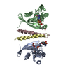 6rirS S: Starting model for refinement C: citing same article ( |
|---|---|
| Similar structure data |


-Links
 PDBj
PDBj


- Assembly
| Deposited unit | 
| ||||||||
|---|---|---|---|---|---|---|---|---|---|
| 1 | 
| ||||||||
| Unit cell |
|
-Components
| #1: Protein | Mass: 21129.160 Da / Num. of mol.: 1 / Mutation: Q67L Source method: isolated from a genetically manipulated source Details: T72 is phosphorylated (TPO) / Source: (gene. exp.) Homo sapiens (human) / Gene: RAB8A, MEL, RAB8 / Production host:  Escherichia coli (E. coli) / References: UniProt: P61006 Escherichia coli (E. coli) / References: UniProt: P61006 |
|---|---|
| #2: Protein | Mass: 6149.065 Da / Num. of mol.: 1 / Fragment: RH2 domain Source method: isolated from a genetically manipulated source Source: (gene. exp.) Homo sapiens (human) / Gene: RILPL2, RLP2 / Production host: Escherichia coli (E. coli) / References: UniProt: Q969X0 |
| #3: Chemical | ChemComp-GTP / Guanosine triphosphate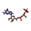  Mass: 523.180 Da / Num. of mol.: 1 / Source method: obtained synthetically / Formula: C10H16N5O14P3 / Comment: GTP, energy-carrying molecule*YM Mass: 523.180 Da / Num. of mol.: 1 / Source method: obtained synthetically / Formula: C10H16N5O14P3 / Comment: GTP, energy-carrying molecule*YM |
| #4: Chemical | ChemComp-MG /   Mass: 24.305 Da / Num. of mol.: 1 / Source method: obtained synthetically / Formula: Mg Mass: 24.305 Da / Num. of mol.: 1 / Source method: obtained synthetically / Formula: Mg |
| #5: Water | ChemComp-HOH / Water Mass: 18.015 Da / Num. of mol.: 69 / Source method: isolated from a natural source / Formula: H2O Mass: 18.015 Da / Num. of mol.: 69 / Source method: isolated from a natural source / Formula: H2O |
| Has ligand of interest | Y |
-Experimental details
-Experiment
| Experiment | Method: X-RAY DIFFRACTION / Number of used crystals: 1 |
|---|
- Sample preparation
| Crystal | Density Matthews: 2.52 Å3/Da / Density % sol: 51.22 % |
|---|---|
| Crystal grow | Temperature: 288 K / Method: vapor diffusion / pH: 7.5 / Details: 150mM DL-Malic acid, 20% PEG3350 |
-Data collection
| Diffraction | Mean temperature: 100 K / Serial crystal experiment: N |
|---|---|
| Diffraction source | Source: SYNCHROTRON / Site: APS  / Beamline: 24-ID-E / Wavelength: 0.97918 Å / Beamline: 24-ID-E / Wavelength: 0.97918 Å |
| Detector | Type: DECTRIS EIGER2 X 16M / Detector: PIXEL / Date: Jul 28, 2019 |
| Radiation | Protocol: SINGLE WAVELENGTH / Monochromatic (M) / Laue (L): M / Scattering type: x-ray |
| Radiation wavelength | Wavelength: 0.97918 Å / Relative weight: 1 |
| Reflection | Resolution: 1.9→46.26 Å / Num. obs: 21414 / % possible obs: 96.02 % / Redundancy: 8.6 % / Biso Wilson estimate: 43.62 Å2 / CC1/2: 0.999 / Rmerge(I) obs: 0.05713 / Rpim(I) all: 0.02056 / Net I/σ(I): 16.79 |
| Reflection shell | Resolution: 1.9→1.968 Å / Redundancy: 7.9 % / Rmerge(I) obs: 4.301 / Mean I/σ(I) obs: 0.35 / Num. unique obs: 1964 / CC1/2: 0.54 / Rpim(I) all: 1.62 / Rrim(I) all: 4.61 / % possible all: 90.3 |
- Processing
| Software |
| |||||||||||||||||||||||||||||||||||||||||||||||||||||||||||||||||||||||||||||||||||||||||||||||||||||||||
|---|---|---|---|---|---|---|---|---|---|---|---|---|---|---|---|---|---|---|---|---|---|---|---|---|---|---|---|---|---|---|---|---|---|---|---|---|---|---|---|---|---|---|---|---|---|---|---|---|---|---|---|---|---|---|---|---|---|---|---|---|---|---|---|---|---|---|---|---|---|---|---|---|---|---|---|---|---|---|---|---|---|---|---|---|---|---|---|---|---|---|---|---|---|---|---|---|---|---|---|---|---|---|---|---|---|---|
| Refinement | Method to determine structure: MOLECULAR REPLACEMENT Starting model: 6rir Resolution: 1.9→46.26 Å / SU ML: 0.36 / Cross valid method: THROUGHOUT / σ(F): 0.23 / Phase error: 39.54 / Stereochemistry target values: ML
| |||||||||||||||||||||||||||||||||||||||||||||||||||||||||||||||||||||||||||||||||||||||||||||||||||||||||
| Solvent computation | Shrinkage radii: 0.9 Å / VDW probe radii: 1.11 Å / Solvent model: FLAT BULK SOLVENT MODEL | |||||||||||||||||||||||||||||||||||||||||||||||||||||||||||||||||||||||||||||||||||||||||||||||||||||||||
| Displacement parameters | Biso max: 142.9 Å2 / Biso mean: 58.0008 Å2 / Biso min: 37.04 Å2 | |||||||||||||||||||||||||||||||||||||||||||||||||||||||||||||||||||||||||||||||||||||||||||||||||||||||||
| Refinement step | Cycle: final / Resolution: 1.9→46.26 Å
| |||||||||||||||||||||||||||||||||||||||||||||||||||||||||||||||||||||||||||||||||||||||||||||||||||||||||
| Refine LS restraints |
| |||||||||||||||||||||||||||||||||||||||||||||||||||||||||||||||||||||||||||||||||||||||||||||||||||||||||
| LS refinement shell | Refine-ID: X-RAY DIFFRACTION / Rfactor Rfree error: 0 / Total num. of bins used: 14
| |||||||||||||||||||||||||||||||||||||||||||||||||||||||||||||||||||||||||||||||||||||||||||||||||||||||||
| Refinement TLS params. | Method: refined / Refine-ID: X-RAY DIFFRACTION
| |||||||||||||||||||||||||||||||||||||||||||||||||||||||||||||||||||||||||||||||||||||||||||||||||||||||||
| Refinement TLS group |
|
