 Movie
Movie Controller

[English] 日本語
 Yorodumi
- PDB-7d7v: Crystal Structure of the Domain1 of NAD+ Riboswitch with nicotina... -
+ Open data

- Basic information
| Entry | Database: PDB / ID: 7d7v | ||||||
|---|---|---|---|---|---|---|---|
| Title | Crystal Structure of the Domain1 of NAD+ Riboswitch with nicotinamide adenine dinucleotide (NAD+) and U1A protein | ||||||
 Components Components |
| ||||||
 Keywords Keywords |  RNA / riboswitch / RNA structure / RNA folding / RNA-ligand interactions / RNA crystallography RNA / riboswitch / RNA structure / RNA folding / RNA-ligand interactions / RNA crystallography | ||||||
| Function / homology |  Function and homology information Function and homology informationU1 snRNP binding / U1 snRNP / U1 snRNA binding / U4/U6 x U5 tri-snRNP complex / mRNA Splicing - Major Pathway / spliceosomal complex / mRNA splicing, via spliceosome / DNA binding / RNA binding / nucleoplasm ...U1 snRNP binding / U1 snRNP / U1 snRNA binding / U4/U6 x U5 tri-snRNP complex / mRNA Splicing - Major Pathway / spliceosomal complex / mRNA splicing, via spliceosome / DNA binding / RNA binding / nucleoplasm / identical protein binding / nucleusSimilarity search - Function | ||||||
| Biological species |  Acidobacterium capsulatum ATCC 51196 (bacteria) Acidobacterium capsulatum ATCC 51196 (bacteria) Homo sapiens (human) Homo sapiens (human) | ||||||
| Method | X-RAY DIFFRACTION / SYNCHROTRON / SAD / Resolution: 2.8 Å | ||||||
 Authors Authors | Chen, H. / Ren, A.M. | ||||||
| Funding support |  China, 1items China, 1items
| ||||||
 Citation Citation | Journal: Nucleic Acids Res. / Year: 2020 Title: Structural distinctions between NAD+ riboswitch domains 1 and 2 determine differential folding and ligand binding. Authors: Chen, H. / Egger, M. / Xu, X. / Flemmich, L. / Krasheninina, O. / Sun, A. / Micura, R. / Ren, A. | ||||||
| History |
|
- Structure visualization
| Structure viewer | Molecule: MolmilJmol/JSmol |
|---|
- Downloads & links
-Download
| PDBx/mmCIF format | 7d7v.cif.gz | 71.9 KB | Display | PDBx/mmCIF format |
|---|---|---|---|---|
| PDB format | pdb7d7v.ent.gz | 48.6 KB | Display | PDB format |
| PDBx/mmJSON format | 7d7v.json.gz | Tree view | PDBx/mmJSON format | |
| Others |  Other downloads Other downloads |
-Validation report
| Arichive directory | https://data.pdbj.org/pub/pdb/validation_reports/d7/7d7vftp://data.pdbj.org/pub/pdb/validation_reports/d7/7d7v | HTTPS FTP |
|---|
-Related structure data
| Related structure data |  7d7wC  7d7xC 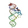 7d7yC 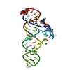 7d7zC 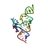 7d81C  7d82C C: citing same article ( |
|---|---|
| Similar structure data |


-Links
 PDBj
PDBj


- Assembly
| Deposited unit | 
| ||||||||
|---|---|---|---|---|---|---|---|---|---|
| 1 |
| ||||||||
| Unit cell |
|
-Components
-RNA chain / Protein , 2 types, 2 molecules AC
| #1: RNA chain | Mass: 18270.900 Da / Num. of mol.: 1 / Mutation: G4del A51del G29C30G31A32 TO AUUGCACUCC Source method: isolated from a genetically manipulated source Source: (gene. exp.) Acidobacterium capsulatum ATCC 51196 (bacteria)Production host: in vitro transcription vector pT7-TP(deltai) (others) |
|---|---|
| #2: Protein | Mass: 10681.421 Da / Num. of mol.: 1 / Mutation: Y27H, Q32R Source method: isolated from a genetically manipulated source Source: (gene. exp.) Homo sapiens (human) / Gene: SNRPA / Production host: bacterium (bacteria) / References: UniProt: P09012 |
-Non-polymers , 4 types, 51 molecules 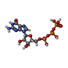


| #3: Chemical | ChemComp-GTP / Guanosine triphosphate Mass: 523.180 Da / Num. of mol.: 1 / Source method: obtained synthetically / Formula: C10H16N5O14P3 / Comment: GTP, energy-carrying molecule*YM Mass: 523.180 Da / Num. of mol.: 1 / Source method: obtained synthetically / Formula: C10H16N5O14P3 / Comment: GTP, energy-carrying molecule*YM | ||||
|---|---|---|---|---|---|
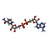
| #4: Chemical | ChemComp-MG /  Mass: 24.305 Da / Num. of mol.: 8 / Source method: obtained synthetically / Formula: Mg / Feature type: SUBJECT OF INVESTIGATION Mass: 24.305 Da / Num. of mol.: 8 / Source method: obtained synthetically / Formula: Mg / Feature type: SUBJECT OF INVESTIGATION#5: Chemical | ChemComp-NAD / | Nicotinamide adenine dinucleotide Mass: 663.425 Da / Num. of mol.: 1 / Source method: obtained synthetically / Formula: C21H27N7O14P2 / Feature type: SUBJECT OF INVESTIGATION / Comment: NAD*YM Mass: 663.425 Da / Num. of mol.: 1 / Source method: obtained synthetically / Formula: C21H27N7O14P2 / Feature type: SUBJECT OF INVESTIGATION / Comment: NAD*YM#6: Water | ChemComp-HOH / | WaterMass: 18.015 Da / Num. of mol.: 41 / Source method: isolated from a natural source / Formula: H2O |
-Details
| Has ligand of interest | Y |
|---|
-Experimental details
-Experiment
| Experiment | Method: X-RAY DIFFRACTION / Number of used crystals: 1 |
|---|
- Sample preparation
| Crystal | Density Matthews: 3.92 Å3/Da / Density % sol: 68.59 % |
|---|---|
| Crystal grow | Temperature: 289 K / Method: vapor diffusion, sitting drop / Details: 0.2 M Mg(OAc)2, 0.1 M C2H6AsNaO2, pH 6.5, 30% MPD |
-Data collection
| Diffraction | Mean temperature: 100 K / Serial crystal experiment: N | |||||||||||||||||||||||||||||||||||||||||||||||||||||||||||||||||||||||||||||||||||||||||||||||||||
|---|---|---|---|---|---|---|---|---|---|---|---|---|---|---|---|---|---|---|---|---|---|---|---|---|---|---|---|---|---|---|---|---|---|---|---|---|---|---|---|---|---|---|---|---|---|---|---|---|---|---|---|---|---|---|---|---|---|---|---|---|---|---|---|---|---|---|---|---|---|---|---|---|---|---|---|---|---|---|---|---|---|---|---|---|---|---|---|---|---|---|---|---|---|---|---|---|---|---|---|---|
| Diffraction source | Source: SYNCHROTRON / Site: SSRF / Beamline: BL18U1 / Wavelength: 0.9793 Å | |||||||||||||||||||||||||||||||||||||||||||||||||||||||||||||||||||||||||||||||||||||||||||||||||||
| Detector | Type: DECTRIS PILATUS3 6M / Detector: PIXEL / Date: Dec 28, 2019 | |||||||||||||||||||||||||||||||||||||||||||||||||||||||||||||||||||||||||||||||||||||||||||||||||||
| Radiation | Protocol: SINGLE WAVELENGTH / Monochromatic (M) / Laue (L): M / Scattering type: x-ray | |||||||||||||||||||||||||||||||||||||||||||||||||||||||||||||||||||||||||||||||||||||||||||||||||||
| Radiation wavelength | Wavelength: 0.9793 Å / Relative weight: 1 | |||||||||||||||||||||||||||||||||||||||||||||||||||||||||||||||||||||||||||||||||||||||||||||||||||
| Reflection | Resolution: 2.8→50 Å / Num. obs: 12481 / % possible obs: 99.6 % / Redundancy: 21.4 % / Rmerge(I) obs: 0.082 / Rpim(I) all: 0.016 / Rrim(I) all: 0.084 / Χ2: 0.842 / Net I/σ(I): 5.1 | |||||||||||||||||||||||||||||||||||||||||||||||||||||||||||||||||||||||||||||||||||||||||||||||||||
| Reflection shell | Diffraction-ID: 1
|
- Processing
| Software |
| ||||||||||||||||||
|---|---|---|---|---|---|---|---|---|---|---|---|---|---|---|---|---|---|---|---|
| Refinement | Method to determine structure: SAD / Resolution: 2.8→50 Å / Cross valid method: THROUGHOUT
| ||||||||||||||||||
| Displacement parameters | Biso max: 139.72 Å2 / Biso mean: 76.524 Å2 / Biso min: 31.73 Å2 | ||||||||||||||||||
| Refinement step | Cycle: LAST / Resolution: 2.8→50 Å
| ||||||||||||||||||
| LS refinement shell | Resolution: 2.8→3.0795 Å
|
